胆管囊肿Bile Duct Cyst · 肝 · 肝胆系统 · NTP ntp-0417
诊断要点:小的胆管囊肿偶尔可见于未处理的对照大鼠,但更常与处理相关。因此,只要存在就应记录并给予严重程度分级。
病变描述
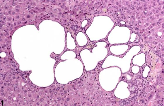
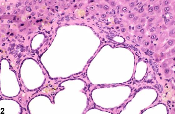
单房和多房性胆管囊肿(biliary cysts)可在老年大鼠和小鼠中自发发生。它们可作为单个扩张的胆管结构、多个相邻的扩张胆管结构(Figure 1、Figure 2)或多房性囊肿(Figure 3)出现。胆管囊肿可伴随肝毒性,并可能与外周或基底部的胶原沉积有关(Figure 4)。它们通常由单层扁平或低立方上皮衬覆,几乎没有活跃增殖的证据。与胆管瘤(cholangiomas)不同,它们不压迫相邻的肝实质。
诊断与分级建议(NTP 共识)
小的胆管囊肿偶尔可见于未处理的对照大鼠,但更常与处理相关。因此,只要存在就应记录并给予严重程度分级。
图谱
英文原文
展开 NTP 官方英文描述与建议
Comment:
Single and multiloculated biliary cysts can occur spontaneously in older rats and mice. They can occur as single dilated biliary structures, multiple adjacent dilated biliary structures ( Figure 1 and Figure 2 ), or a multiloculated cyst ( Figure 3 ). Biliary cysts may accompany hepatic toxicity and may be associated with peripheral or sub-basilar collagen deposition ( Figure 4 ). They are typically lined by a single layer of flattened or low cuboidal epithelium ( Figure 4 ), with little evidence of active proliferation. In contrast to cholangiomas, they do not compress the adjacent hepatic parenchyma.
Recommendation:
Small biliary cysts may occasionally be seen in untreated rats but are more often associated with treatment. Consequently, they should be documented whenever present and given a severity grade.
来源与引用
原始条目:https://ntp.niehs.nih.gov/atlas/nnl/hepatobiliary-system/liver/BileDuct-Cyst
图谱总目录:NTP Nonneoplastic Lesion Atlas。 内容属美国联邦政府作品(公有领域),可引用;引用时请标注来源。
建议引用格式:NTP Nonneoplastic Lesion Atlas. Bile Duct Cyst (liver). LabSmart 中文版:
https://www.labsmart.cn/ntp/liver-bile-duct-cyst.html
中文图谱总页(可展开对照全部 595 条):https://www.labsmart.cn/ntp-atlas.html#liver-bile-duct-cyst