枯否细胞增生Kupffer Cell Hyperplasia · 肝 · 肝胆系统 · NTP ntp-0404
诊断要点:该组织学改变在未处理啮齿类动物中罕见,一旦出现即应作出诊断并分级。应在病理学描述中详细说明其形态学特征及组织分布。
病变描述
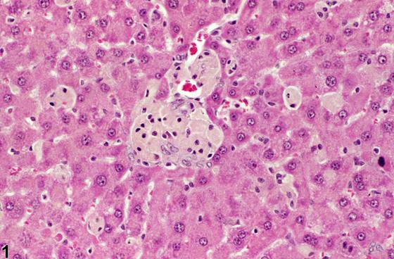
Kupffer细胞肥大与增生通常同时发生,可见于摄取外源性物质、受试物或代谢产物之后,其中任一情况均可能使细胞质着色为色素性。Kupffer细胞增生与肥大亦可发生于炎症状态下。增大的Kupffer细胞可形成片状或小结节状。
诊断与分级建议(NTP 共识)
该组织学改变在未处理啮齿类动物中罕见,一旦出现即应作出诊断并分级。应在病理学描述中详细说明其形态学特征及组织分布。由于肥大是这些细胞吞噬活性的结果,通常无需单独作出肥大的诊断。若色素沉着显著,可单独作出色素沉着的诊断,并附严重程度的定性分级。
图谱
英文原文
展开 NTP 官方英文描述与建议
Comment:
Kupffer cell hypertrophy and hyperplasia usually occur together and can be seen following uptake of foreign material, test agent, or metabolite, any of which may impart a pigment to the cytoplasm. Kupffer hyperplasia and hypertrophy may also occur in inflammatory conditions. The enlarged Kupffer cells may form sheets or small nodules.
Recommendation:
This tissue change is rare in untreated rodents and should be diagnosed and graded whenever present. A description of the morphologic features and tissue distribution should be detailed in the pathology narrative. Because hypertrophy is a consequence of the phagocytic activity of these cells, a separate diagnosis of hypertrophy is generally not necessary. If pigmentation is prominent, a separate diagnosis of pigmentation, along with a qualitative grading of severity, may be appropriate.
来源与引用
原始条目:https://ntp.niehs.nih.gov/atlas/nnl/hepatobiliary-system/liver/KupfferCell-Hyperplasia
图谱总目录:NTP Nonneoplastic Lesion Atlas。 内容属美国联邦政府作品(公有领域),可引用;引用时请标注来源。
建议引用格式:NTP Nonneoplastic Lesion Atlas. Kupffer Cell Hyperplasia (liver). LabSmart 中文版:
https://www.labsmart.cn/ntp/liver-kupffer-cell-hyperplasia.html
中文图谱总页(可展开对照全部 595 条):https://www.labsmart.cn/ntp-atlas.html#liver-kupffer-cell-hyperplasia