胆管纤维化Cholangiofibrosis · 肝 · 肝胆系统 · NTP ntp-0419
诊断要点:这种肝脏炎症反应应予以记录并给出严重程度分级,严重程度取决于肝脏受累范围。上述所有特征均包括在胆管纤维化的诊断中,不应单独诊断,除非它们明确代表独立的病变过程。
病变描述
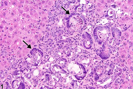
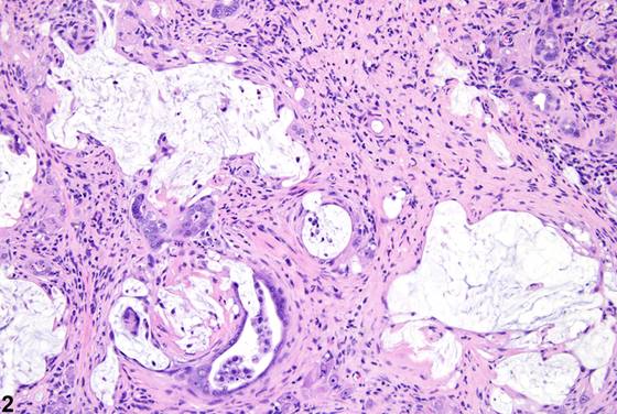
这种慢性炎症过程最初伴有卵圆细胞增生和胆管增生,增生的胆管扩张。胆管内容物包括黏液、坏死碎片和脱落的上皮细胞(Figure 3)。胆道结构常由深染的立方至柱状细胞和杯状细胞(肠上皮化生)不完全衬覆(新月形胆管;Figure 1,箭头)。随着病变进展,出现胆管周围纤维化和混合性炎症细胞浸润(Figure 1、Figure 2)。肝叶的大面积区域可能受累,病变通过硬化性结缔组织的形成而逐渐消退,同时保留部分胆道腺泡和残留的黏液池。胆管纤维化可能类似胆管癌,但可通过后者更为显著的胆管上皮改变(例如上皮细胞多层或堆积以及多形性)加以区分。然而,上皮细胞异型性和核分裂象增多可见于这两种病变,因此无助于区分胆管纤维化与胆管癌。一些文献指出,明显的胆管扩张和胆管上皮的肠上皮化生并非胆管癌的显著特征。
诊断与分级建议(NTP 共识)
这种肝脏炎症反应应予以记录并给出严重程度分级,严重程度取决于肝脏受累范围。上述所有特征均包括在胆管纤维化的诊断中,不应单独诊断,除非它们明确代表独立的病变过程。相关链接:肝脏 - 炎症
图谱
英文原文
展开 NTP 官方英文描述与建议
Comment:
This chronic inflammatory process is initially associated with oval cell proliferation and bile duct hyperplasia with dilation of proliferating bile ducts. Bile duct contents include mucus, necrotic debris, and desquamated epithelial cells ( Figure 3 ). Biliary structures are frequently incompletely lined (crescent-shaped bile ducts; Figure 1 , arrows) by hyperchromatic cuboidal to columnar cells and goblet cells (intestinal metaplasia). As the lesion progresses, there is peribiliary fibrosis and a mixed inflammatory cell infiltrate ( Figure 1 and Figure 2 ). Large areas of hepatic lobe may be affected, with progressive resolution of lesions by development of sclerotic connective tissue, with retention of some biliary glandular acini and residual pools of mucus. Cholangiofibrosis may resemble cholangiocarcinoma but may be differentiated by the latter’s more prominent biliary epithelial changes (e.g., multiple layers or piling up of epithelial cells and pleomorphism). Epithelial cell atypia and increased mitotic figures may be present in either lesion, however, and therefore are not useful in differentiating cholangiofibrosis from cholangiocarcinoma. Some references indicate that significant bile duct dilation and intestinal metaplasia of the biliary epithelium are not prominent features of cholangiocarcinoma.
Recommendation:
This inflammatory hepatic response should be documented and given a severity grade, with severity dependent on the extent of liver involvement. All the features described above are included in the diagnosis of cholangiofibrosis and should not be diagnosed separately unless they clearly represent separate processes. Related Links: Liver - Inflammation
来源与引用
原始条目:https://ntp.niehs.nih.gov/atlas/nnl/hepatobiliary-system/liver/Cholangiofibrosis
图谱总目录:NTP Nonneoplastic Lesion Atlas。 内容属美国联邦政府作品(公有领域),可引用;引用时请标注来源。
建议引用格式:NTP Nonneoplastic Lesion Atlas. Cholangiofibrosis (liver). LabSmart 中文版:
https://www.labsmart.cn/ntp/liver-cholangiofibrosis.html
中文图谱总页(可展开对照全部 595 条):https://www.labsmart.cn/ntp-atlas.html#liver-cholangiofibrosis