脂肪变性Fatty Change · 肝 · 肝胆系统 · NTP ntp-0420
诊断要点:脂肪变的形态学表现具有足够的特征性,无需确认性特殊染色或其他程序即可作出诊断。只要存在,就应记录肝细胞脂肪变的诊断。
病变描述
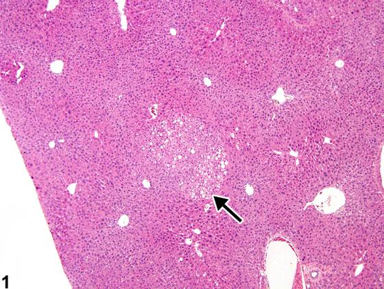
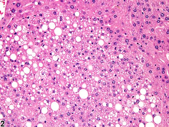
脂肪变(fatty change)可为局灶性(focal)(Figure 1,箭头;Figure 2)或弥漫性(diffuse)(Figure 3、Figure 4、Figure 5、Figure 6),并可为大泡性(macrovesicular)(Figure 3、Figure 4)或小泡性(microvesicular)(Figure 5、Figure 6)。大泡性脂肪变常与代谢紊乱相关,且通常易于逆转,而小泡性脂肪变更可能是毒性的反映,可能涉及线粒体功能障碍。Figure 1、Figure 2代表局灶性病变,其中空泡化肝细胞的形态学特征同时符合大泡性和小泡性脂肪变。局灶性脂肪变可为自发性病变,且在某些品系中可能比其他品系更常见,而弥漫性或带状(如小叶中心性或门管区周围性)脂肪变更可能与受试物相关。Figure 3、Figure 4中的细胞质空泡化为弥漫性,由肝细胞内大小不一的单个或双个细胞质空泡组成。其形态学特征符合大泡性脂肪变,伴有受累肝细胞核受压缩并被推移至周边。相比之下,Figure 5、Figure 6代表小泡性脂肪变,在门管区周围和小叶中部区域反应更为显著。细胞质改变由充满细胞质的多个小空泡组成,无明显肝细胞核压缩或移位。肝窦可被肥大的肝细胞压迫而塌陷。Figure 7、Figure 8代表大泡性脂肪变与肝细胞内糖原蓄积的混合。可见糖原与大泡性及小泡性脂肪变的混合形式。磷脂沉积症(phospholipidosis)是肝细胞细胞质空泡化的另一种形式,与暴露于阳离子两亲性外源化学物相关,需特殊染色和/或电子显微镜检查才能确诊。
诊断与分级建议(NTP 共识)
脂肪变的形态学表现具有足够的特征性,无需确认性特殊染色或其他程序即可作出诊断。只要存在,就应记录肝细胞脂肪变的诊断。由于未处理或溶媒对照动物中可能发生一定程度的脂肪变,推荐进行严重程度分级以记录潜在的受试物相关效应。由于局灶性(focal)脂肪变可能为自发性,当病变为局灶性时,诊断中应包含分布修饰词“局灶性”。当病变为弥漫性(diffuse)时,不应使用分布修饰词(即,当诊断中未指定分布时,假定病变为弥漫性)。脂肪变的形态学特征(如大泡性、小泡性或两者兼有)应在病理学描述中予以描述。其他特征性表现,如分布模式,也应在病理学描述中予以描述。脂肪变常伴有其他细胞质改变,如糖原蓄积或耗竭,且细胞质形态学上不同类型的改变可能难以区分。在这些情况下,适合作出肝细胞细胞质空泡化的诊断并附严重程度分级,但应在病理学描述中详尽描述肝细胞细胞质改变的形态学。病理学描述还应包括病理学家关于细胞质空泡内容的意见。相关链接:肝脏——张力性脂质沉积症(Liver - Tension Lipidosis)
图谱
英文原文
展开 NTP 官方英文描述与建议
Comment:
Fatty change can be focal ( Figure 1 , arrow; Figure 2 ) or diffuse ( Figure 3 , Figure 4 , Figure 5 , and Figure 6 ) and macrovesicular ( Figure 3 and Figure 4 ) or microvesicular ( Figure 5 and Figure 6 ). Macrovesicular fatty change is often associated with metabolic disturbances and is generally readily reversible, whereas microvesicular fatty change is more likely a reflection of toxicity, possibly involving mitochondrial disturbances. Figures Figure 1 and Figure 2 represent a focal lesion in which the morphologic features of the vacuolated hepatocytes are consistent with both macrovesicular and microvesicular fatty change. Focal fatty change can be a spontaneous lesion and may be more common in some strains than others, whereas diffuse or zonal (e.g., centrilobular or periportal) fatty change is more likely to be treatment related. The cytoplasmic vacuolization in Figures Figure 3 and Figure 4 is diffuse and consists of both large and small single or double cytoplasmic vacuoles within hepatocytes. The morphologic features are consistent with macrovesicular fatty change, with compression and displacement of the nuclei to the periphery of affected hepatocytes. In contrast, Figure 5 and Figure 6 are representative of microvesicular fatty change, with a more prominent response in periportal and midlobular areas. The cytoplasmic change consists of multiple small vacuoles filling the cytoplasm without prominent compression or displacement of hepatocyte nuclei. Sinusoids may be collapsed by the hypertrophic hepatocytes. Figure 7 and Figure 8 represent a mixture of both macrovesicular fatty change and glycogen accumulation in hepatocytes. Mixtures of glycogen and both macrovesicular and microvesicular fatty change may be seen. Phospholipidosis is another form of hepatocyte cytoplasmic vacuolization that is associated with exposure to cationic amphophilic xenobiotics and requires special staining and/or electron microscopy for definitive confirmation.
Recommendation:
The morphologic appearance of fatty change is distinctive enough to allow its diagnosis in the absence of confirmatory special stains or other procedures. Whenever present, diagnosis of hepatocellular fatty change should be recorded. Since some degree of fatty change may occur in untreated or vehicle controls, severity grading is recommended to document potential treatment-related effects. Because focal fatty change may be spontaneous, the distribution modifier “focal” should be included in the diagnosis when the lesion is focal. When the lesion is diffuse, no distribution modifier should be used (i.e., the lesion is assumed to be diffuse when the distribution is not specified in the diagnosis). The morphologic features of the fatty change (e.g., macrovesicular, microvesicular, or both) should be described in the pathology narrative. Other distinctive features, such as the pattern of distribution, should also be described in the pathology narrative. Fatty change is frequently accompanied by other cytoplasmic alterations, such as glycogen accumulation or depletion, and it can be difficult to distinguish the different types of alterations in cytoplasmic morphology. In those situations, a diagnosis of hepatocyte cytoplasmic vacuolization is appropriate, along with a severity grade, but the morphology of the cytoplasmic changes in the hepatocytes should be thoroughly described in the pathology narrative. The pathology narrative should also include the pathologist’s opinions regarding the contents of the cytoplasmic vacuoles. Related Links: Liver - Tension Lipidosis
来源与引用
原始条目:https://ntp.niehs.nih.gov/atlas/nnl/hepatobiliary-system/liver/FattyChange
图谱总目录:NTP Nonneoplastic Lesion Atlas。 内容属美国联邦政府作品(公有领域),可引用;引用时请标注来源。
建议引用格式:NTP Nonneoplastic Lesion Atlas. Fatty Change (liver). LabSmart 中文版:
https://www.labsmart.cn/ntp/liver-fatty-change.html
中文图谱总页(可展开对照全部 595 条):https://www.labsmart.cn/ntp-atlas.html#liver-fatty-change