灶Focus · 肝 · 肝胆系统 · NTP ntp-0406
诊断要点:由于灶是推定的癌前病变,只要存在即应诊断。NTP目前的建议是记录所有灶并将其分类为亚型(嗜碱性、嗜酸性、透明细胞或混合型)。
病变描述
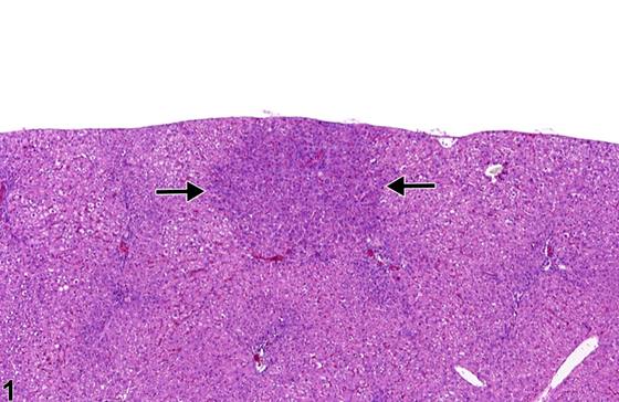
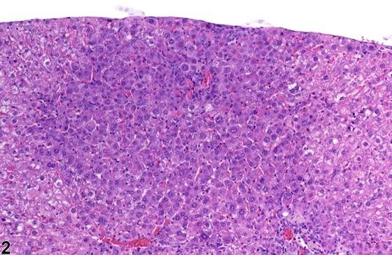
细胞改变灶(foci of cellular alteration)在NTP研究中简称为“灶”(foci),可在大鼠和小鼠中自发发生,但也可能由受试物诱导产生。其大小范围从小于一个肝小叶到数个肝小叶不等。灶是推定的癌前病变(presumptive preneoplastic lesions),其表现可从几乎难以察觉至细胞形态学和染色特性上界限分明的病变不等。灶通常与周围肝实质界限不清地融合,且不压迫周围肝实质,但可能发生轻微压迫。灶在慢性研究中相对常见,但在90天研究中不常见。根据苏木精-伊红(hematoxylin and eosin)染色特性和细胞质特征对其进行亚分类。嗜碱性灶(basophilic foci)主要被苏木精着色(Figure 1,箭头;Figure 2)。在大鼠中已鉴定出虎斑型变体(tigroid variant),但被视为嗜碱性灶的一个亚分类。嗜酸性灶(eosinophilic foci)的染色通常比周围肝细胞更具嗜酸性,且常由比邻近正常实质更大的肝细胞组成(Figure 3,箭头;Figure 4)。透明细胞灶(clear cell foci)在中央定位的细胞核周围的肝细胞胞质中具有空泡。这些透明空泡代表在固定和处理过程中溶解流失的糖原。透明细胞灶易于识别,因为它们在胞质嗜酸性更为均匀的周围实质背景下显得突出(Figure 5、Figure 6)。某些文献讨论了空泡细胞灶(vacuolated cell foci),但这些现在被诊断为局灶性(focal) fatty change,不再被视为灶。当不存在单一优势表型时,诊断为混合细胞灶(mixed cell focus)。Figure 7、Figure 8展示了一个由透明细胞、空泡细胞和双染性细胞(amphophilic cells)组成的混合细胞灶,其中无单一细胞类型占灶细胞的80%以上。Figure 9、Figure 10展示了一个混合灶,由外周的嗜碱性细胞环围绕内部的透明细胞核心组成,各占现存细胞类型的相等比例。
诊断与分级建议(NTP 共识)
由于灶是推定的癌前病变,只要存在即应诊断。NTP目前的建议是记录所有灶并将其分类为亚型(嗜碱性、嗜酸性、透明细胞或混合型)。灶不需要严重程度分级,除非病理学家认为有必要突出研究组间的差异。相关链接:肝脏 - 肝细胞 - 糖原蓄积与耗竭(Liver - Hepatocyte - Glycogen Accumulation and Depletion)
图谱
英文原文
展开 NTP 官方英文描述与建议
Comment:
Foci of cellular alteration, diagnosed simply as “foci” in NTP studies, can occur spontaneously in rats and mice but may also be induced by treatment. They range from less than a lobule to several lobules in size. Foci are presumptive preneoplastic lesions that can vary from barely perceptible to cytomorphologically and tinctorially discrete lesions. Foci typically blend imperceptibly with, and do not compress, surrounding hepatic parenchyma, though minimal compression may occur. Foci are relatively common in chronic studies but uncommon in 90-day studies. They are subclassified based on hematoxylin and eosin tinctorial properties and cytoplasmic features. Basophilic foci predominantly stain with hematoxylin ( Figure 1 , arrows; Figure 2 ). A tigroid variant has been identified in rats but is considered a subclassification of basophilic focus. Eosinophilic foci typically stain more eosinophilic than surrounding hepatocytes and often consist of hepatocytes that are larger than the adjacent normal parenchyma ( Figure 3 , arrows; Figure 4 ). Clear cell foci have empty spaces in hepatocyte cytoplasm surrounding centrally localized nuclei. The clear spaces represent glycogen dissolved out during fixation and processing. Clear cell foci are readily identified because they stand out against surrounding parenchyma that has a more uniformly eosinophilic cytoplasm ( Figure 5 and Figure 6 ). Some texts discuss vacuolated cell foci, but these are now diagnosed as focal fatty change and are not considered foci. Mixed cell focus is diagnosed when there is no single predominant phenotype. Figure 7 and Figure 8 represent a mixed cell focus composed of clear cells, vacuolated cells, and amphophilic cells with no single cell type constituting more than 80% of the focus cells. Figure 9 and Figure 10 present a mixed focus composed of an outer rim of basophilic cells surrounding an inner core of clear cells, each comprising an equal proportion of the cell types present.
Recommendation:
Because foci are presumptive preneoplastic lesions, they should be diagnosed whenever present. The current NTP recommendation is to document all foci and to classify these into subtypes (basophilic, eosinophilic, clear cell, or mixed). A severity grade is not required for foci unless the pathologist feels it is necessary to highlight differences among the study groups. Related Links: Liver - Hepatocyte - Glycogen Accumulation and Depletion
来源与引用
原始条目:https://ntp.niehs.nih.gov/atlas/nnl/hepatobiliary-system/liver/Focus
图谱总目录:NTP Nonneoplastic Lesion Atlas。 内容属美国联邦政府作品(公有领域),可引用;引用时请标注来源。
建议引用格式:NTP Nonneoplastic Lesion Atlas. Focus (liver). LabSmart 中文版:
https://www.labsmart.cn/ntp/liver-focus.html
中文图谱总页(可展开对照全部 595 条):https://www.labsmart.cn/ntp-atlas.html#liver-focus