肝细胞肥大Hepatocyte Hypertrophy · 肝 · 肝胆系统 · NTP ntp-0414
诊断要点:肝细胞肥大只要存在即应诊断并分级。轻微病例可能难以诊断,在许多情况下,需要与同期对照进行比较以确认诊断。
病变描述
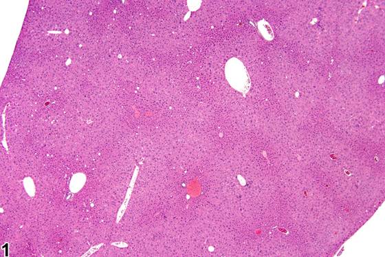
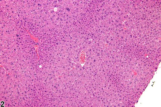
肝细胞肥大(hepatocyte hypertrophy)是一种细胞学改变,其诊断依据是与同期对照肝脏相比,肝细胞体积出现可观察到的增大。当肝细胞肥大呈现常见的中央小叶(centrilobular)分布模式时,最为显而易见;当其为全小叶性(panlobular)时,与同期对照的比较可提供诊断性确认。当病变为轻度时,肝细胞肥大的检出可能较为困难,但在低倍放大下其识别会更为容易。肝细胞肥大通常与继发于某些外源性化学物(xenobiotics)暴露的微粒体酶诱导相关(Figure 1、Figure 2、Figure 3)。其最常累及中央小叶肝细胞,具体取决于外源性化学物及给药剂量,尽管肥大可扩展至肝小叶中部或变为全小叶性。也可能以门管区周围(periportal)肝细胞受累为主,但必须注意不要将门管区周围肥大与导致中央小叶肝细胞皱缩的过程(例如糖原耗竭)相混淆。Figure 4 为同一研究中正常的对照肝脏,用于与 Figure 3 进行比较。取决于所涉及的具体外源性化学物,肥大肝细胞的胞质可呈苍白、毛玻璃样外观,或呈颗粒状且嗜酸性增强,尤其是在过氧化物酶体增殖剂(peroxisome proliferators)暴露后(Figure 5)。Figure 6 为同期对照肝脏,用于与 Figure 5 进行比较。长期暴露于某些非遗传毒性肝脏毒物时,可见伴有核增大(karyomegaly)和多核肝细胞的显著肥大。Figure 7 中可见一狭窄的中央小叶区肝细胞肥大(Figure 8 为更高倍放大)。增大肝细胞中细颗粒状嗜酸性胞质与过氧化物酶体增殖时所见的相似,但在本例中,导致肝细胞肥大的细胞学改变被认为是透明变性(hyaline change)。在极端形式下,透明变性可形成明确的胞质内包涵体,有时称为Mallory小体(Mallory bodies)。Figure 9 为 Figure 5 中央静脉区的高倍放大,显示肝细胞凋亡(箭头)和胆色素潴留(箭)。由于增大的肝细胞压迫肝窦导致血流减少,可发生肝细胞变性和细胞死亡。
诊断与分级建议(NTP 共识)
肝细胞肥大只要存在即应诊断并分级。轻微病例可能难以诊断,在许多情况下,需要与同期对照进行比较以确认诊断。如果怀疑肥大但不明确,则适宜对切片进行盲法评估,将相关剂量组与对照进行比较。当改变处于临界状态时,伴随的肝脏重量增加可为诊断提供一定的支持。在可能的情况下,肝细胞肥大的诊断应附以分布模式限定,并给予定性严重程度分级。如果肝细胞增大是由于胞质空泡或包涵体的存在所致,则不应诊断肝细胞肥大;而应记录胞质空泡化或胞质内包涵体,除非有其他确凿的肥大证据。
图谱
英文原文
展开 NTP 官方英文描述与建议
Comment:
Hepatocyte hypertrophy is a form of cytologic alteration that is diagnosed based on an observable increased size of hepatocytes compared with concurrent control liver. It is most readily apparent when it has the commonly occurring centrilobular distribution pattern; when it is panlobular, comparison with concurrent controls can provide diagnostic confirmation. When mild, detection of hepatocyte hypertrophy may be difficult, but its identification is facilitated at low magnification. Hepatocyte hypertrophy is commonly associated with microsomal enzyme induction secondary to exposure to certain xenobiotics ( Figure 1 , Figure 2 , and Figure 3 ). It most frequently affects centrilobular hepatocytes, depending upon the xenobiotic and the dose administered, although the hypertrophy can extend into the middle of the hepatic lobule or become panlobular. It is also possible for periportal hepatocytes to be primarily affected, though care must be taken not to confuse periportal hypertrophy with processes that result in shrinkage of centrilobular hepatocytes (e.g., glycogen depletion). Figure 4 is a normal control liver from the same study for comparison with Figure 3 . Depending upon the specific xenobiotic responsible, hypertrophic hepatocyte cytoplasm may have a pale, ground glass appearance or be granular and intensely eosinophilic, especially following exposure to peroxisome proliferators ( Figure 5 ). Figure 6 is a concurrent control liver for comparison with Figure 5 . Marked hypertrophy with associated karyomegaly and multinucleated hepatocytes may be seen with chronic exposure to some nongenotoxic hepatic toxicants. A narrow centrilobular zone of hepatocyte hypertrophy is present in Figure 7 (higher magnification in Figure 8 ). The finely granular eosinophilic cytoplasm in the enlarged hepatocytes is similar to that seen with peroxisome proliferation, but in this case the cytologic alteration responsible for the hepatocyte hypertrophy was considered hyaline change. In extreme forms, hyaline change can form distinct cytoplasmic inclusions, sometimes referred to as Mallory bodies. Figure 9 is a high magnification of the central vein area of Figure 5 and shows apoptosis of hepatocytes (arrowhead) and retention of bile pigment (arrows). Hepatocyte degeneration and cell death can occur because of reduced blood flow secondary to sinusoidal compression by enlarged hepatocytes.
Recommendation:
Hepatocyte hypertrophy should be diagnosed and graded whenever present. Subtle cases may be difficult to diagnose, and in many instances, comparison with concurrent controls is necessary to confirm the diagnosis. If hypertrophy is suspected but not clear-cut, then a blind evaluation of the slides comparing the relevant dose groups with controls is appropriate. Accompanying increases in liver weight may provide some comfort in diagnosis when the change is marginal. When possible, the diagnosis of hepatocyte hypertrophy should be qualified with a distribution pattern and given a qualitative severity grade. If the hepatocytes are enlarged due to the presence of cytoplasmic vacuoles or inclusions, hypertrophy should not be diagnosed; rather, the cytoplasmic vacuolization or cytoplasmic inclusions should be recorded, unless there is other, definitive evidence of hypertrophy.
来源与引用
原始条目:https://ntp.niehs.nih.gov/atlas/nnl/hepatobiliary-system/liver/Hepatocyte-Hypertrophy
图谱总目录:NTP Nonneoplastic Lesion Atlas。 内容属美国联邦政府作品(公有领域),可引用;引用时请标注来源。
建议引用格式:NTP Nonneoplastic Lesion Atlas. Hepatocyte Hypertrophy (liver). LabSmart 中文版:
https://www.labsmart.cn/ntp/liver-hepatocyte-hypertrophy.html
中文图谱总页(可展开对照全部 595 条):https://www.labsmart.cn/ntp-atlas.html#liver-hepatocyte-hypertrophy