肝细胞核分裂增多Hepatocyte - Increased Mitosis · 肝 · 肝胆系统 · NTP ntp-0410
诊断要点:核分裂频率增高并不常见;无论何时出现都应记录,并给予定性严重程度分级。
病变描述
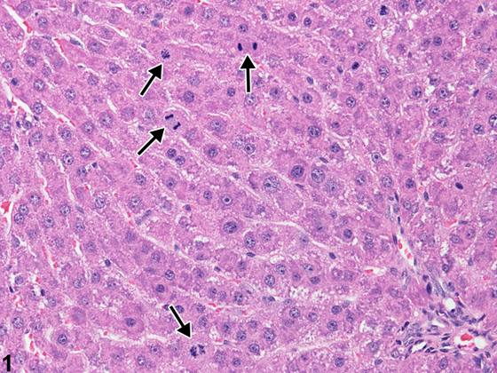
在早期生长阶段、妊娠等生理状态下,或在对其他部位荷瘤的啮齿类动物中,可见高的核分裂频率。虽然在正常肝脏中可见偶见的核分裂,但每10个高倍视野中发现超过一或两个核分裂对成年啮齿类动物来说并不典型。在本例中,高的核分裂频率(Figure 1,箭头)是肝毒性物质处理后继发于肝细胞丢失的修复反应。
诊断与分级建议(NTP 共识)
核分裂频率增高并不常见;无论何时出现都应记录,并给予定性严重程度分级。
图谱
英文原文
展开 NTP 官方英文描述与建议
Comment:
A high mitotic frequency can be seen during phases of early growth, during physiologic conditions such as pregnancy, or in rodents bearing tumors at other sites. While occasional mitoses can be seen in a normal liver, finding more than one or two mitoses per 10 high-power fields is not typical for adult rodents. In this example, the high frequency of mitosis ( Figure 1 , arrows) is a repair response following hepatocyte loss secondary to treatment with a hepatotoxicant.
Recommendation:
An increased frequency of mitosis is unusual; it should be documented whenever present and given a qualitative severity grade.
来源与引用
原始条目:https://ntp.niehs.nih.gov/atlas/nnl/hepatobiliary-system/liver/Hepatocyte%E2%80%93IncreasedMitosis
图谱总目录:NTP Nonneoplastic Lesion Atlas。 内容属美国联邦政府作品(公有领域),可引用;引用时请标注来源。
建议引用格式:NTP Nonneoplastic Lesion Atlas. Hepatocyte - Increased Mitosis (liver). LabSmart 中文版:
https://www.labsmart.cn/ntp/liver-hepatocyte-increased-mitosis.html
中文图谱总页(可展开对照全部 595 条):https://www.labsmart.cn/ntp-atlas.html#liver-hepatocyte-increased-mitosis