炎症Inflammation · 肝 · 肝胆系统 · NTP ntp-0407
诊断要点:形态学上与上述局灶性(focal)炎症描述一致的炎症性病变应记录为“炎症,局灶性”,无论是否与受试物相关。
病变描述
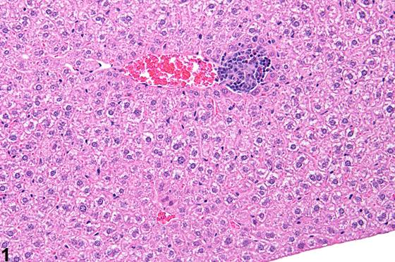
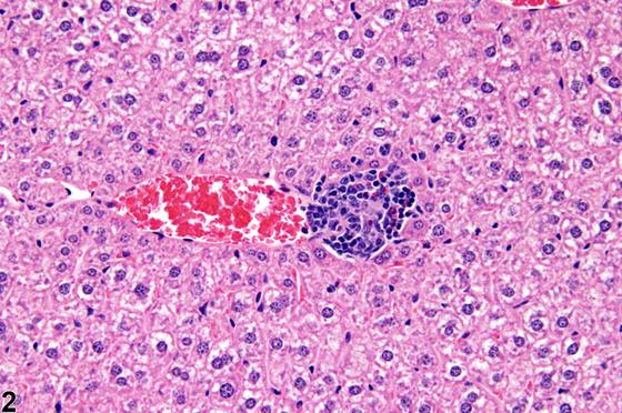
炎症,局灶性(focal)是指单个或多个、局灶性、随机分布的炎症细胞聚集灶,在肝脏中作为背景性病变可见,且在老年小鼠和大鼠中比在年轻动物中更为常见。然而,这些炎症细胞灶可在亚慢性研究的啮齿类动物肝脏中自发出现(Figure 1、Figure 2)。浸润细胞以淋巴细胞为主,但可能包含较少量的中性粒细胞和/或巨噬细胞。炎症细胞聚集灶可能伴有肝细胞坏死的证据(Figure 3、Figure 4)。这些灶的大小可能不一(Figure 6、Figure 7),但通常肉眼不明显。这些病变可能在雌性动物中更为常见(Harada et al. 1996),并可能与感染性病原体有关(Hall et al. 1992)。最常见的原因是未知的。如果在标准组织学切片中仅见一或两个炎症灶,则严重程度分级为1+(极轻微(minimal))。与同期对照相比,外源性化学物暴露也可能增加这些病变的严重程度和/或数量。局灶性炎症是啮齿类动物毒性研究中肝脏最常见的炎症性病变,是慢性炎症的一种形式。在慢性炎症中,以淋巴细胞为主。慢性活动性炎症同样以淋巴细胞为主,但也包含相当数量的中性粒细胞。两种病变均可能含有巨噬细胞。然而,只有当病变在某种定性方式上与上述“炎症,局灶性”的描述不同时,才可使用“慢性炎症”或“慢性活动性炎症”这一术语。其他较少见的炎症形式包括急性、化脓性和肉芽肿性炎症。在急性炎症中,中性粒细胞是主要的浸润细胞,尽管也可能存在较少的巨噬细胞和淋巴细胞,以及水肿或充血的表现。在化脓性炎症中,中性粒细胞同样是主要的浸润细胞类型,但它们聚集在一起,且许多已变性。渗出物中还可能存在来自 resident 细胞群和浸润白细胞的细胞碎片,以及含有纤维蛋白的蛋白性液体、少量巨噬细胞、偶见的淋巴细胞或浆细胞,以及可能的感染性病原体。肉眼上,这些病变以脓液的存在为特征。渗出物周围组织可能含有成纤维细胞、纤维结缔组织和混合性炎症细胞,取决于病变的慢性程度。肉芽肿性炎症是另一种形式的慢性炎症,其诊断要求存在大量聚集的、大的活化巨噬细胞、上皮样巨噬细胞和/或多核巨细胞。“细胞浸润”一词曾被用于描述炎症细胞的存在而无炎症过程的其他证据(例如水肿、细胞坏死或变性,或血管损伤的证据)。此类炎症细胞浸润通常相当小,可能不值得诊断。如果足够显著而值得记录,白细胞聚集应诊断为“炎症”。偶尔,肝脏中可见非白细胞性细胞(如肥大细胞)的浸润;这些应诊断为“细胞浸润”。
诊断与分级建议(NTP 共识)
形态学上与上述局灶性(focal)炎症描述一致的炎症性病变应记录为“炎症,局灶性”,无论是否与受试物相关。严重程度分级应反映组间任何与受试物相关的差异。对照组中一或两个小的局灶性炎症细胞聚集灶,在受试物组中无任何加重、增多或减少的情况下,被视为背景性病变。如果在特定研究中未记录,病理学叙述应说明其偶尔可见且研究各组间无任何差异。如果在研究中记录,则每次观察到时均应一致地记录和分级。由于该病变与衰老相关,其在28天或90天研究中的出现可能值得记录。如果存在的炎症性病变在某种定性方式上(形态学、分布等)与上述局灶性炎症的描述不同,则可使用上述其他诊断术语之一。然而,由于局灶性炎症是如此常见的病变,且可能与其他类型的炎症同时存在,这些病变必须在叙述中详细描述,以便能与局灶性炎症明确区分。“炎症”一词应用于指白细胞,但当肝脏中存在非白细胞性细胞(如肥大细胞)时,可使用“浸润,细胞性”。所有这些病变应根据肝脏受累范围进行分级。形态学特征如分布模式可在病理学叙述中呈现。任何相关变化,如肝细胞变性、坏死、色素沉着或血管变化,如果这些相关病变的严重程度值得,可作为独立病变诊断。相关链接:肝脏 - 胆管纤维化(cholangiofibrosis)
图谱
英文原文
展开 NTP 官方英文描述与建议
Comment:
Inflammation, focal is the term used to denote single or multiple, focal, randomly distributed aggregates of inflammatory cells that are seen in the liver as a background lesion and are more frequent in aging mice and rats than in young animals. However, these foci of inflammatory cells can occur spontaneously in livers of rodents in prechronic studies ( Figure 1 and Figure 2 ). The infiltrating cells are predominantly lymphocytes but may include fewer numbers of neutrophils and/or macrophages. Inflammatory cell aggregates may be accompanied by evidence of hepatocellular necrosis ( Figure 3 and Figure 4 ). These foci may vary in size ( Figure 6 and Figure 7 ) but typically are not grossly apparent. These lesions may be more common in females (Harada et al. 1996) and may be related to infectious agents (Hall et al. 1992). Most often the cause is unknown. If only one or two foci of inflammation are seen in the standard histologic sections, the severity grade would be 1+ (minimal). Xenobiotic exposure may also increase the severity and/or number of these lesions compared with concurrent controls. Focal inflammation is the most frequently seen inflammatory lesion of the liver in rodent toxicity studies and is a form of chronic inflammation. In chronic inflammation, lymphocytes predominate. Chronic active inflammation predominately involves lymphocytes as well but also includes a significant number of neutrophils. Both lesions may contain macrophages. However, the term “chronic inflammation” or “chronic active inflammation” may be used only if the lesion differs in some qualitative way from the above description of “inflammation, focal.” Other less common forms of inflammation include acute, suppurative, and granulomatous inflammation. In acute inflammation , neutrophils are the predominant infiltrating cell, though fewer macrophages and lymphocytes may also be present, as well as evidence of edema or hyperemia. In suppurative inflammation , neutrophils are also the predominant infiltrating cell type but they are aggregated, and many are degenerate. Cell debris from both the resident cell populations and infiltrating leukocytes may also be present in the exudate, as well as proteinaceous fluid containing fibrin, a few macrophages, occasional lymphocytes or plasma cells, and possibly an infectious agent. Grossly, these lesions would be characterized by the presence of pus. The tissue surrounding the exudate may contain fibroblasts, fibrous connective tissue, and mixed inflammatory cells, depending on the chronicity of the lesion. Granulomatous inflammation is another form of chronic inflammation whose diagnosis requires the presence of a significant number of aggregated, large, activated macrophages, epithelioid macrophages, and/or multinucleated giant cells. The term “cellular infiltrate” has been used to describe the presence of inflammatory cells without other evidence of an inflammatory process (e.g., edema, necrosis or degeneration of cells, or evidence of vascular injury). Such infiltrates of inflammatory cells are generally quite small and may not warrant diagnosis. If significant enough to warrant documentation, accumulations of leukocytes should be diagnosed as “inflammation.” Occasionally, infiltrates of nonleukocytic cells such as mast cells are seen in the liver; these should be diagnosed as “cellular infiltrate.”
Recommendation:
Inflammatory lesions that are morphologically consistent with the above description of focal inflammation should be recorded as “inflammation, focal” whether or not they are related to treatment. The severity grades should reflect any treatment-related differences between groups. One or two small focal inflammatory cell aggregates in controls in the absence of any exacerbation, increase, or decrease in treated groups is considered a background lesion. If it is not documented in a specific study, the pathology narrative should indicate that it was occasionally seen without any differences among study groups. If it is documented in a study, it should be consistently recorded and graded whenever seen. Because this lesion is associated with aging, its occurrence in 28- or 90-day studies may warrant documentation. If an inflammatory lesion is present that differs in some qualitative manner (morphology, distribution, etc.) from the above description of focal inflammation, one of the other diagnostic terms described above may be used. However, because focal inflammation is such a common lesion and may be present concurrently with another type of inflammation, these lesions must be thoroughly described in the narrative so that they may be clearly differentiated from focal inflammation. The term “inflammation” should be used in reference to leukocytes, but “infiltration, cellular” may be used when nonleukocytic cells are present in the liver (e.g., mast cells). All of these lesions should be graded based on the extent of liver involvement. Morphologic features such as the distribution pattern can be presented in the pathology narrative. Any associated changes, such as hepatocyte degeneration, necrosis, pigmentation, or vascular changes, may be diagnosed as a separate lesion if warranted by the severity of these associated lesions. Related Links: Liver - Cholangiofibrosis
来源与引用
原始条目:https://ntp.niehs.nih.gov/atlas/nnl/hepatobiliary-system/liver/Inflammation
图谱总目录:NTP Nonneoplastic Lesion Atlas。 内容属美国联邦政府作品(公有领域),可引用;引用时请标注来源。
建议引用格式:NTP Nonneoplastic Lesion Atlas. Inflammation (liver). LabSmart 中文版:
https://www.labsmart.cn/ntp/liver-inflammation.html
中文图谱总页(可展开对照全部 595 条):https://www.labsmart.cn/ntp-atlas.html#liver-inflammation