结晶Crystals · 肺 · 呼吸系统 · NTP ntp-0112
诊断要点:细胞内和气道内游离的晶体只要存在,均应诊断为 Lung - Crystals 并进行分级。
病变描述
在NTP研究中,肺内晶体最常见于采用颗粒物吸入研究的小鼠(Figure 1、Figure 2、Figure 3),或与肿瘤相关。这些晶体常与引起显著肺泡蛋白沉积症的药物同时出现。晶体呈嗜酸性(eosinophilic)且为针状或纺锤形。部分较大的晶体可能呈 elongated rectangles 或“砖块状”。它们通常存在于肺泡腔内,并伴有组织细胞浸润,或在更严重的病例中伴有肉芽肿性炎症。晶体常被大的巨噬细胞包围,这些巨噬细胞可能为多核,部分较小的晶体可能位于巨噬细胞和多核巨细胞的细胞质内。部分巨噬细胞可能含有大量小晶体(Figure 3)。通常,间质或肺泡腔内存在数量不等的其他炎症细胞(淋巴细胞、浆细胞、中性粒细胞)。血红蛋白晶体偶尔见于出血区域(Figure 4),常伴有炎症。此类晶体最常见于大鼠,因为大鼠血红蛋白比其他物种的血红蛋白更易结晶。在其他物种中,只有结构异常的血红蛋白才会在体内结晶。血红蛋白晶体通常呈矩形,与嗜酸性结晶性肺炎的嗜酸性晶体不同,其形成于细胞外空间,尽管较小的血红蛋白晶体可能被肺泡巨噬细胞吞噬。其颜色通常类似红细胞,且通常与外渗的红细胞在空间上相关。嗜酸性结晶性肺炎是一种发生于小鼠的特发性疾病。它可以是散发性背景病变,严重程度从 轻微(mild) 和亚临床到 severe 不等,严重时可导致死亡。它可见于某些基因工程小鼠(例如 p47 phox-/- 小鼠)或由自然发生的突变培育而成的小鼠品系(例如 Ptpn6 me 裸鼠)。易感品系包括 C57BL/6 和 129Sv 小鼠。嗜酸性结晶性肺炎的发病机制尚不清楚。在易感品系中,它曾与嗜酸性炎症相关,例如由寄生虫和真菌感染引起的嗜酸性炎症,尽管与晶体相关的主要细胞类型是肺泡巨噬细胞。这些晶体对 anti-Ym1 protein 抗体(又称 T 淋巴细胞来源的嗜酸性粒细胞趋化因子)呈免疫反应性。嗜酸性结晶性肺炎在组织学上与颗粒物吸入相关的晶体(及炎症)相似,但与任何处理均无关联,并且也可见于对照小鼠。在啮齿类动物肺中已鉴定出其他类型的晶体。大鼠亚慢性暴露于氯法齐明(一种抗麻风药物)会导致红橙色晶体沉积于许多组织,包括肺。在 F344 大鼠中长期经饮食给予曲酸可产生含有晶体的肺部微肉芽肿。C57BL/6 小鼠感染新型隐球菌(Cryptococcus neoformans)后产生杆状晶体结构,这些结构可见于与酵母细胞相关或游离于宿主细胞质中。尽管这些晶体在形态上类似于小鼠嗜酸性结晶性肺炎中所见的晶体,但推测这些晶体是宿主蛋白与隐球菌荚膜多糖相互作用的产物。
诊断与分级建议(NTP 共识)
细胞内和气道内游离的晶体只要存在,均应诊断为 Lung - Crystals 并进行分级。病理学描述中应包含对晶体的详尽描述,包括大小、形状、颜色、位置以及是细胞内还是细胞外(以及所见于何种细胞类型)。伴随的炎症应单独诊断并分级。如果晶体是与肿瘤相关的炎症性病变的一部分,则无需诊断,但应在病理学描述中予以描述。如果病理学家认为晶体是小鼠嗜酸性结晶性肺炎的一部分,也应在病理学描述中注明。
图谱
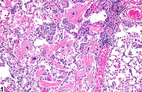
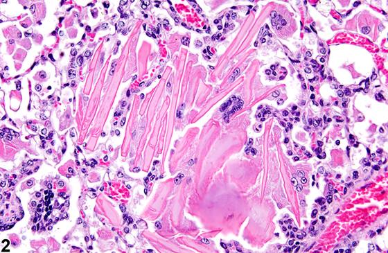
英文原文
展开 NTP 官方英文描述与建议
Comment:
In NTP studies, intrapulmonary crystals are most often seen in mice in inhalation studies with particulates ( Figure 1 , Figure 2 , and Figure 3 ) or associated with neoplasms. The crystals are frequently seen with agents that cause significant alveolar proteinosis. The crystals are eosinophilic and acicular, or spindle shaped. Some larger crystals may be elongated rectangles, or "brick shaped." They are usually present in the alveolar spaces and are associated with histiocytic infiltrates or, in more severe cases, granulomatous inflammation. The crystals are often surrounded by large macrophages, which may be multinucleated, and some smaller crystals may be within the cytoplasm of the macrophages and multinucleated giant cells. Some macrophages may contain numerous small crystals ( Figure 3 ). Typically, variable numbers of other inflammatory cells (lymphocytes, plasma cells, neutrophils) are present in the interstitium or in the alveolar spaces. Hemoglobin crystals are occasionally seen in areas of hemorrhage ( Figure 4 ), often associated with inflammation. This type of crystal is most commonly seen in rats because rat hemoglobin crystallizes more readily than does hemoglobin in other species. Only structurally abnormal hemoglobin crystallizes in vivo in other species. Hemoglobin crystals are typically rectangular and, as opposed to the eosinophilic crystals of eosinophilic crystalline pneumonia, form in the extracellular space, though smaller hemoglobin crystals may be phagocytosed by alveolar macrophages. They typically resemble red blood cells in color and are usually spatially associated with extravasated erythrocytes. Eosinophilic crystalline pneumonia is an idiopathic disease that occurs in mice. It can be a sporadic background lesion, with the severity varying from mild and subclinical to severe, in which case it may cause death. It may be seen in some genetically engineered mice (e.g., p47 phox-/- mice) or mouse strains developed from naturally occurring mutations (e.g., Ptpn6 me motheaten mice). Susceptible strains include C57BL/6 and 129Sv mice. The pathogenesis of eosinophilic crystalline pneumonia is unclear. It has been associated with eosinophilic inflammation in susceptible strains, such as that caused by parasitic and fungal infections, though the main cell type associated with the crystals is the alveolar macrophage. The crystals are immunoreactive to anti-Ym1 protein antibodies (a.k.a. T-lymphocyte-derived eosinophil chemotactic factor). Eosinophilic crystalline pneumonia is histologically similar to the crystals (and inflammation) associated with inhalation of particulates but does not occur with any relation to treatment and would also be seen in control mice. Other types of crystals have been identified in rodent lungs. Subchronic exposure of rats to clofazimine, an antileprotic drug, leads to deposition of reddish orange crystals in many tissues, including lung. Chronic dietary administration of kojic acid in F344 rats produced pulmonary microgranulomas containing crystals. Cryptococcus neoformans infection in C57BL/6 mice generated rod-like crystalline structures found associated with yeast cells or free in host cell cytoplasm. Though these crystals appear morphologically similar to those seen in murine eosinophilic crystalline pneumonia, these crystals are presumed to be the product of interaction of a host protein and a capsular cryptococcal polysaccharide.
Recommendation:
Crystals both intracellular and free within airspaces should be diagnosed as Lung - Crystals and graded whenever present. A thorough description of the crystals, including size, shape, color, location, and whether they are intra- or extracellular (and the cell types in which they are found), should be included in the pathology narrative. Accompanying inflammation should be diagnosed and graded separately. If they are part of an inflammatory lesion associated with a neoplasm, they need not be diagnosed but should be described in the pathology narrative. If, in the opinion of the pathologist, the crystals are part of murine eosinophilic crystalline pneumonia, this should also be noted in the pathology narrative.
来源与引用
原始条目:https://ntp.niehs.nih.gov/atlas/nnl/respiratory-system/lung/Crystals
图谱总目录:NTP Nonneoplastic Lesion Atlas。 内容属美国联邦政府作品(公有领域),可引用;引用时请标注来源。
建议引用格式:NTP Nonneoplastic Lesion Atlas. Crystals (lung). LabSmart 中文版:
https://www.labsmart.cn/ntp/lung-crystals.html
中文图谱总页(可展开对照全部 595 条):https://www.labsmart.cn/ntp-atlas.html#lung-crystals