再生Regeneration · 肺 · 呼吸系统 · NTP ntp-0097
诊断要点:肺 - 再生(Lung - Regeneration):只要存在,且同时存在坏死或变性证据,即应诊断。
病变描述
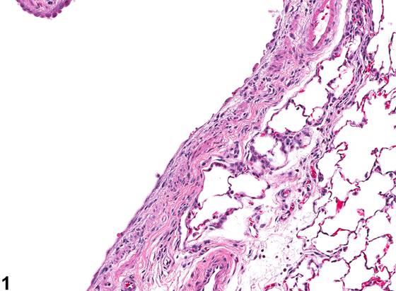
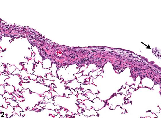
再生(regeneration)是指被覆上皮表面(即气道和肺泡)裸露部分得以修复的过程。一旦上皮发生坏死或溃疡,邻近的存活上皮细胞即取代丢失的细胞。在气道中,该过程始于邻近上皮细胞的伸长,这些细胞变扁平,并迁移越过创面覆盖基底膜(Figure 1、Figure 2)。在后期阶段,上皮开始增殖以取代丢失的细胞,使上皮衬里恢复正常状态。在肺泡中,II型肺泡上皮细胞(type II pneumocytes)是祖细胞,经历上述过程以取代丢失的I型肺泡上皮细胞。在某些情况下,再生的上皮可能超过正常上皮细胞数量而变为增生(hyperplastic)(Figure 3)。在此类情况下,在缺乏可识别的坏死或溃疡时,可能难以确定增生是源于再生还是对受试物暴露的原发性反应。因此,“再生”一词保留用于存在上皮损伤证据的病例。再生的其他特征可能包括与细胞增殖相符的特征,如核增大(karyomegaly)和细胞增大(cytomegaly)。此外,增殖细胞可能具有轻度非典型外观,因此病理学家可能需要区分再生与细胞非典型性(cellular atypia)。
诊断与分级建议(NTP 共识)
肺 - 再生(Lung - Regeneration):只要存在,且同时存在坏死或变性证据,即应诊断。如果上皮细胞数量超出正常范围,则应诊断增生(hyperplasia),无论其是受试物暴露的直接效应还是过度再生,但增生的发病机制应在病理学描述中讨论。诊断中应包含部位修饰词(如支气管、细支气管、肺泡),以指示病变位置。再生不应进行分级。为传达病变的严重程度,必须同时诊断并分级坏死或变性(见 Lung - Epithelium - Degeneration 和 Lung - Epithelium - Necrosis)。其他相关病变,如炎症,应单独诊断。如果病理学家判断再生上皮表现出显著的非典型特征,则应诊断并分级 Lung - Atypia, Cellular。相关链接:Lung - Atypia, Cellular;Lung - Epithelium - Degeneration;Lung - Epithelium - Necrosis
图谱
英文原文
展开 NTP 官方英文描述与建议
Comment:
Regeneration is the process whereby denuded portions of epithelium-lined surfaces (i.e., airways and alveoli) are repaired. Once the epithelium becomes necrotic or ulcerated, the adjacent, viable epithelial cells replace the lost cells. In the airways, the process begins with elongation of the adjacent epithelial cells, which become flattened, and migration of these cells across the wound to cover the basement membrane ( Figure 1 and Figure 2 ). In later stages, the epithelium begins to proliferate to replace the lost cells to return the epithelial lining to its normal state. In the alveoli, the type II pneumocytes are the progenitor cells and undergo these processes to replace lost alveolar type I cells. In some cases, the regenerating epithelium may overshoot the normal number of epithelial cells and become hyperplastic ( Figure 3 ). In such cases, in the absence of recognizable necrosis or ulceration, it may be difficult to determine whether the hyperplasia is due to regeneration or is a primary response to test article exposure. For this reason, the term "regeneration" is reserved for those cases where there is evidence of epithelial damage. Other features of regeneration may include characteristics consistent with cellular proliferation, such as karyomegaly and cytomegaly. Additionally, the proliferating cells may have a slightly atypical appearance, so the pathologist may need to distinguish between regeneration and cellular atypia.
Recommendation:
Lung - Regeneration should be diagnosed whenever present and evidence of necrosis or degeneration is also present. If the number of epithelial cells exceeds that which would be considered within normal limits, hyperplasia should be diagnosed, regardless of whether it is a direct effect of test article exposure or excessive regeneration, but the pathogenesis of the hyperplasia should be discussed in the pathology narrative. A site modifier (e.g., bronchus, bronchiole, alveolus) should be included in the diagnosis to indicate the location of the lesion. Regeneration should not be graded. To convey the severity of the lesion, either necrosis or degeneration must be diagnosed concurrently and graded (see Lung - Epithelium - Degeneration and Lung - Epithelium - Necrosis ). Other associated lesions, such as inflammation, should be diagnosed separately. If the pathologist judges that the regenerating epithelium exhibits significant features of atypia, then Lung - Atypia, Cellular should be diagnosed and graded. Related Links: Lung - Atypia, Cellular Lung - Epithelium - Degeneration Lung - Epithelium - Necrosis
来源与引用
原始条目:https://ntp.niehs.nih.gov/atlas/nnl/respiratory-system/lung/Regeneration
图谱总目录:NTP Nonneoplastic Lesion Atlas。 内容属美国联邦政府作品(公有领域),可引用;引用时请标注来源。
建议引用格式:NTP Nonneoplastic Lesion Atlas. Regeneration (lung). LabSmart 中文版:
https://www.labsmart.cn/ntp/lung-regeneration.html
中文图谱总页(可展开对照全部 595 条):https://www.labsmart.cn/ntp-atlas.html#lung-regeneration