上皮变性Epithelium Degeneration · 肺 · 呼吸系统 · NTP ntp-0095
诊断要点:肺,上皮——变性只要存在就应诊断并分级。诊断中应包含部位修饰词以指示病变在肺内的位置(例如肺泡、细支气管),因为毒性损伤可优先靶向特定部位。
病变描述
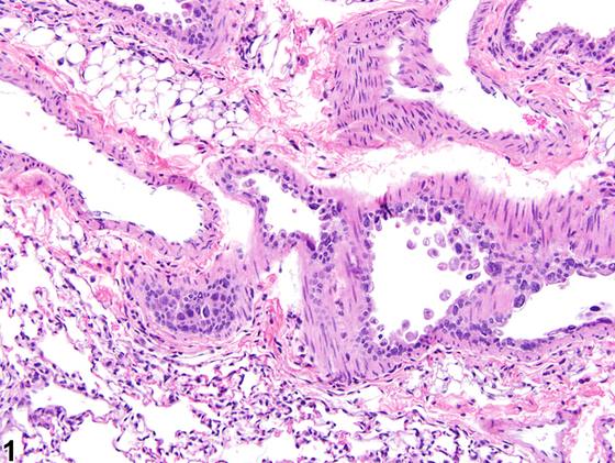
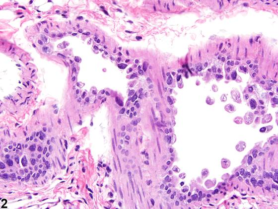
变性(Figure 1、Figure 2、Figure 3、Figure 4)和坏死被认为是细胞损伤连续过程的一部分,其中变性代表可逆性细胞损伤,坏死代表不可逆性细胞损伤。可逆性细胞损伤(变性)的光镜特征包括细胞肿胀、胞质空泡化、核周透明间隙、胞质泡形成、Clara 细胞正常顶端泡丢失以及纤毛丢失。在某些情况下,存活细胞从上皮表面脱落以及上皮内散在细胞的核浓缩(pyknosis)和细胞皱缩,提示单个细胞即将死亡,可被解释为上皮变性,因为这可能与上皮表面的可逆性损伤相一致,且可能缺乏明确的坏死证据。坏死的光镜特征包括核浓缩、核碎裂或核溶解、细胞肿胀、细胞细节丢失、细胞碎裂以及胞质嗜酸性增强(其中胞质常呈均质外观)。毒性损伤所致气道被覆上皮细胞坏死,常以坏死细胞或细胞碎片脱落入管腔为特征。其他病变常伴随坏死和变性出现,如炎症和出血。变性病变的解剖位置可能因受试物的理化性质或特定细胞类型对受试物的易感性而有所不同。终末细支气管和肺泡管上皮(即中心腺泡区)及肺泡特别易受损伤,这是由于表面积大且 I 型肺泡细胞脆弱、Clara 细胞中 P450 酶的代谢活性以及黏液层通常较薄。
诊断与分级建议(NTP 共识)
肺,上皮——变性只要存在就应诊断并分级。诊断中应包含部位修饰词以指示病变在肺内的位置(例如肺泡、细支气管),因为毒性损伤可优先靶向特定部位。变性与坏死在形态学上存在显著重叠,因此病理学家在诊断这些病变时需要运用其最佳判断。如果变性继发于另一过程,如严重炎症,则最好诊断主要过程并在叙述中描述变性。如果变性和并发的相关病变(例如炎症)均显著,病理学家可选择记录两者并分别分级。同样,病理学家在决定是否单独诊断变性时需要运用其判断。
图谱
英文原文
展开 NTP 官方英文描述与建议
Comment:
Degeneration ( Figure 1 , Figure 2 , Figure 3 , and Figure 4 ) and necrosis are considered to be parts of the continuum of cell damage, with degeneration representing reversible cell damage and necrosis representing irreversible cell damage. The light microscopic hallmarks of reversible cell damage (degeneration) include cellular swelling, cytoplasmic vacuolation, perinuclear clear spaces, formation of cytoplasmic blebs, loss of normal apical blebs from Clara cells, and loss of cilia. In some cases, detachment of viable cells from the epithelial surface and nuclear condensation (pyknosis) and cellular shrinkage of scattered cells within the epithelium, suggestive of imminent death of individual cells, may be interpreted as epithelial degeneration because it may be consistent with reversible damage to an epithelial surface and evidence of outright necrosis may be lacking. The light microscopic features of necrosis include nuclear pyknosis, karyorrhexis, or karyolysis, cell swelling, loss of cellular detail, cell fragmentation, and cytoplasmic hypereosinophilia (in which the cytoplasm often has a homogeneous appearance). Necrosis of the epithelial cells lining the airways as a result of toxic injury is often characterized by sloughing of necrotic cells or cellular debris into the lumen. Other lesions often accompany necrosis and degeneration, such as inflammation and hemorrhage. The anatomic location of degenerative lesions may vary due to the physicochemical properties of the test agent or the susceptibility of a particular cell type to the test agent. The epithelium of the terminal bronchioles and alveolar ducts (i.e., the centriacinar region) and alveoli are particularly susceptible to injury due to the large surface area and fragility of the alveolar type I cells, the metabolic activity of P450 enzymes in Clara cells, and the generally thinner mucous layer.
Recommendation:
Lung, Epithelium - Degeneration should be diagnosed and graded whenever present. A site modifier should be included in the diagnosis to indicate the location of the lesion within the lung (e.g., alveolus, bronchiole) since toxic insults can preferentially target specific sites. There is significant overlap, morphologically, between degeneration and necrosis, so the pathologist will need to use his or her best judgment when diagnosing these lesions. If the degeneration is secondary to another process, such as severe inflammation, it is preferable to diagnose the major process and to describe the degeneration in the narrative. If degeneration and a concurrent, related lesion (e.g., inflammation) are both prominent, the pathologist may choose to record both lesions and grade them separately. Again, the pathologist will need to use his or her judgment in deciding whether or not to diagnose the degeneration separately.
来源与引用
原始条目:https://ntp.niehs.nih.gov/atlas/nnl/respiratory-system/lung/Epithelium-Degeneration
图谱总目录:NTP Nonneoplastic Lesion Atlas。 内容属美国联邦政府作品(公有领域),可引用;引用时请标注来源。
建议引用格式:NTP Nonneoplastic Lesion Atlas. Epithelium Degeneration (lung). LabSmart 中文版:
https://www.labsmart.cn/ntp/lung-epithelium-degeneration.html
中文图谱总页(可展开对照全部 595 条):https://www.labsmart.cn/ntp-atlas.html#lung-epithelium-degeneration