角化性囊肿Keratinizing Cyst · 肺 · 呼吸系统 · NTP ntp-0118
诊断要点:肺 - 角化囊肿只要存在就应诊断,但不应进行分级。然而,如果囊肿数量和/或大小呈剂量相关性增加,且病理学家认为必须对囊肿进行分级才能充分描述该病变与染毒的关系,则可以对囊肿进行分级。
病变描述
角化囊肿(keratinizing cyst)典型见于大鼠吸入颗粒性物质(如滑石粉、二氧化钛)后,也可见于经口灌胃暴露于二噁英类化合物的大鼠。尚未在小鼠或仓鼠中报道。角化囊肿由囊壁和囊腔构成,囊壁为分化良好的鳞状上皮细胞,呈有序成熟,围绕囊腔,腔内含亮嗜酸性、同心圆层状角蛋白(Figure 1、Figure 2、Figure 3、Figure 4)。囊肿可见于任何肺叶,常位于胸膜下。囊肿可变得相当大,直径可达数厘米,并取代或压迫正常肺实质。构成囊肿的鳞状上皮细胞被认为起源于衬覆肺泡和/或细支气管上皮的化生。角化囊肿也曾被称为“鳞状囊肿(squamous cyst)”和“表皮囊肿(epidermal cyst)”。角化囊肿被认为是大鼠角化性病变连续谱的一部分,该谱还包括肺泡上皮鳞状化生、囊性角化上皮瘤和鳞状细胞癌。囊性角化上皮瘤与角化囊肿的鉴别在于上皮瘤的壁更厚、更复杂,外缘更粗糙(囊肿外缘更光滑),且鳞状上皮细胞可能缺乏有序成熟,核分裂象可能增多。鳞状细胞癌可能与囊性角化上皮瘤相似,但表现出恶性证据(侵袭、细胞异型性、细胞极性丧失等)。大鼠中也曾报道仅由纤维组织衬覆的先天性囊性腔隙,其与角化囊肿的鉴别在于缺乏鳞状上皮。
诊断与分级建议(NTP 共识)
肺 - 角化囊肿只要存在就应诊断,但不应进行分级。然而,如果囊肿数量和/或大小呈剂量相关性增加,且病理学家认为必须对囊肿进行分级才能充分描述该病变与染毒的关系,则可以对囊肿进行分级。如果对囊肿进行分级,则必须在病理学叙述中完整描述分级方案。病理学叙述中还应包括不同剂量组中囊肿的肺叶分布及大致数量和大小的描述。相关病变,如炎症,应单独诊断。
图谱
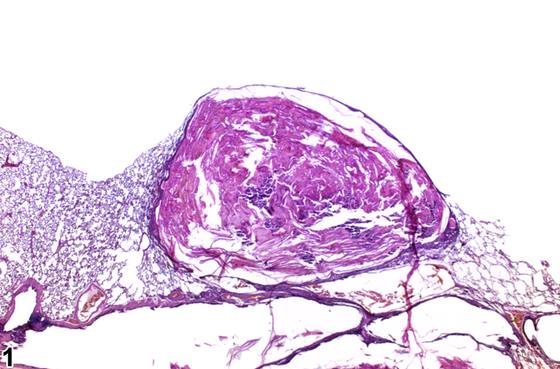
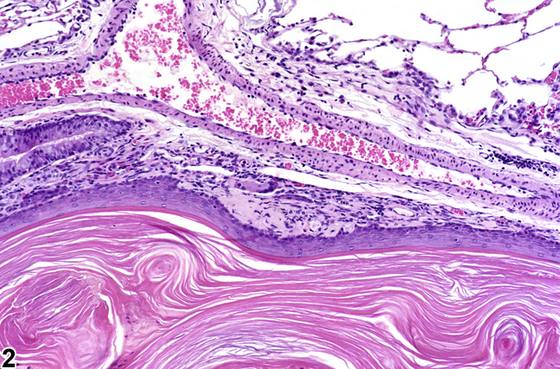
英文原文
展开 NTP 官方英文描述与建议
Comment:
Keratinizing cysts are typically seen in rats after inhalation exposure to particulate material (e.g., talc, titanium dioxide), though they are also seen in rats exposed by gavage to dioxin-like compounds. They have not been reported in mice or hamsters. Keratinizing cysts consist of a cyst wall composed of well-differentiated squamous epithelial cells undergoing orderly maturation surrounding a lumen containing brightly eosinophilic, concentrically laminated keratin ( Figure 1 , Figure 2 , Figure 3 , and Figure 4 ). Cysts can be observed in any lung lobe and are often subpleural. Cysts can become quite large, up to several centimeters in diameter, and replace or compress the normal lung parenchyma. The squamous epithelial cells comprising the cyst are considered to originate from metaplasia of epithelium lining the alveoli and/or bronchioles. Keratinizing cysts have also been called "squamous cysts" and "epidermal cysts." Keratinizing cysts are thought to be part of a continuum of keratinizing lesions seen in rats that also includes squamous metaplasia of the alveolar epithelium, cystic keratinizing epithelioma, and squamous cell carcinoma. Cystic keratinizing epitheliomas can be differentiated from keratinizing cysts by virtue of the thicker, more complex wall of epitheliomas, which has a rougher outer margin (cysts have a smoother outer margin) and may lack orderly maturation of the squamous epithelial cells and may have increased mitotic figures. Squamous cell carcinomas may appear similar to cystic keratinizing epitheliomas but show evidence of malignancy (invasion, cellular atypia, loss of cellular polarity, etc.). Congenital cystic spaces lined only by fibrous tissue have also been reported in the rat, and these are distinguishable from keratinizing cysts by the lack of squamous epithelium.
Recommendation:
Lung - keratinizing cyst should be diagnosed whenever present but should not be graded. However, if a dose-related increase in the number and/or size is present, and the pathologist feels that grading the cysts is necessary to fully characterize the relationship of the lesion to treatment, the cysts may be graded. If the cysts are graded, the grading scheme must be fully described in the pathology narrative. A description of the lobar distribution and approximate number and size of the cysts in the different dose groups should also be included in the pathology narrative. Associated lesions, such as inflammation, should be diagnosed separately.
来源与引用
原始条目:https://ntp.niehs.nih.gov/atlas/nnl/respiratory-system/lung/KeratinizingCyst
图谱总目录:NTP Nonneoplastic Lesion Atlas。 内容属美国联邦政府作品(公有领域),可引用;引用时请标注来源。
建议引用格式:NTP Nonneoplastic Lesion Atlas. Keratinizing Cyst (lung). LabSmart 中文版:
https://www.labsmart.cn/ntp/lung-keratinizing-cyst.html
中文图谱总页(可展开对照全部 595 条):https://www.labsmart.cn/ntp-atlas.html#lung-keratinizing-cyst