杯状细胞化生Metaplasia Goblet Cell · 肺 · 呼吸系统 · NTP ntp-0103
诊断要点:该病变的首选术语为肺-化生,杯状细胞(Lung - Metaplasia, Goblet cell)。
病变描述
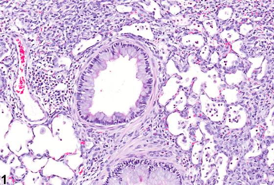
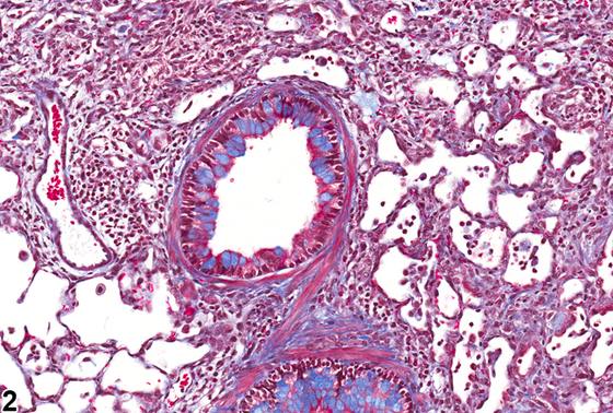
关于该病变的恰当术语(化生与增生)一直存在诸多争议,但由于杯状细胞(goblet cell)通常不存在于较小气道的上皮中,且在上气道中通常不可见,因此在NTP研究中应将其诊断为杯状细胞化生(goblet cell metaplasia)。大鼠和小鼠肺内杯状(黏液)细胞数量极少。在正常条件下,主支气管(mainstem bronchi)、一级支气管(primary bronchi)和叶支气管(lobar bronchi)(约至第六级气道)上皮细胞中不足1-2%为杯状细胞。在段支气管(segmental bronchi)(第7-11级)或终末细支气管(terminal bronchioles)中尚未发现杯状细胞。杯状细胞化生的特征为支气管或细支气管衬覆上皮中出现杯状细胞(数量超过正常范围;见上文)(Figure 1、Figure 2、Figure 3)。在大鼠和小鼠中,杯状细胞化生可与感染性病原体或吸入性刺激物暴露相关。该病变常与炎症同时出现,较少与纤维化同时出现,但也可见于无上述病变的情况下。黏液可用Alcian blue法染色,有助于识别该病变。黏液在Mason三色染色法(Mason's trichrome staining method)中也呈蓝色(Figure 2),但该染色通常不用于识别杯状细胞,首选Alcian blue。
诊断与分级建议(NTP 共识)
该病变的首选术语为肺-化生,杯状细胞(Lung - Metaplasia, Goblet cell)。该病变存在时应记录并分级。化生的部位(即支气管或细支气管)应在诊断中作为部位修饰词标明。若病变存在于多个部位,可省略部位修饰词,并在病理学描述中描述各部位。相关病变,如炎症和细支气管上皮增生,应分别诊断并分级。
图谱
英文原文
展开 NTP 官方英文描述与建议
Comment:
There has been much debate regarding the proper term for this lesion (metaplasia vs. hyperplasia), but since goblet cells are not normally present in the epithelium of the smaller airways and are not normally visible in the upper airways, it should be diagnosed as goblet cell metaplasia in NTP studies. Rats and mice have very low numbers of goblet (mucous) cells in their lungs. Under normal conditions, <1-2% of the epithelial cells of the mainstem bronchi, primary bronchi, and lobar bronchi (up to approximately the sixth-generation airway) are goblet cells. No goblet cells have been identified in the segmental bronchi (generations 7-11) or in the terminal bronchioles. Goblet cell metaplasia is characterized by the presence of goblet cells (above the normal number; see above) in the epithelium lining the bronchi or bronchioles ( Figure 1 , Figure 2 , and Figure 3 ). Goblet cell metaplasia may occur in association with infectious agents or exposure to inhaled irritants in both rats and mice. It is frequently seen concurrently with inflammation and, less frequently, fibrosis, though it may be seen in the absence of these lesions. Mucus can be stained with the Alcian blue method, which can facilitate the identification of this lesion. Mucus also stains blue with the Mason''s trichrome staining method ( Figure 2 ), but this stain is not typically used to identify goblet cells, and Alcian blue is preferred.
Recommendation:
The preferred terminology for this lesion is Lung - Metaplasia, Goblet cell. This lesion should be recorded and graded when present. The location of the metaplasia (i.e., bronchus or bronchiole) should be indicated in the diagnosis as a site modifier. If the lesion is present in more than one location, the site modifier may be omitted and the locations described in the pathology narrative. Associated lesions, such as inflammation and bronchiolar epithelial hyperplasia, should be diagnosed and graded separately.
来源与引用
原始条目:https://ntp.niehs.nih.gov/atlas/nnl/respiratory-system/lung/MetaplasiaGobletCell
图谱总目录:NTP Nonneoplastic Lesion Atlas。 内容属美国联邦政府作品(公有领域),可引用;引用时请标注来源。
建议引用格式:NTP Nonneoplastic Lesion Atlas. Metaplasia Goblet Cell (lung). LabSmart 中文版:
https://www.labsmart.cn/ntp/lung-metaplasia-goblet-cell.html
中文图谱总页(可展开对照全部 595 条):https://www.labsmart.cn/ntp-atlas.html#lung-metaplasia-goblet-cell