鳞状化生Metaplasia Squamous · 肺 · 呼吸系统 · NTP ntp-0120
诊断要点:肺——化生,鳞状:只要存在即应诊断并赋予严重程度分级。诊断中应包含部位修饰词(即肺泡、细支气管或支气管)以指示病变的位置。
病变描述
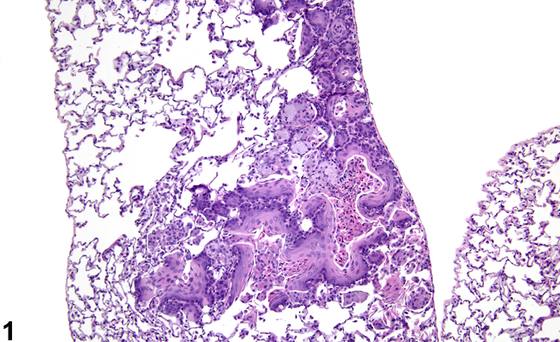
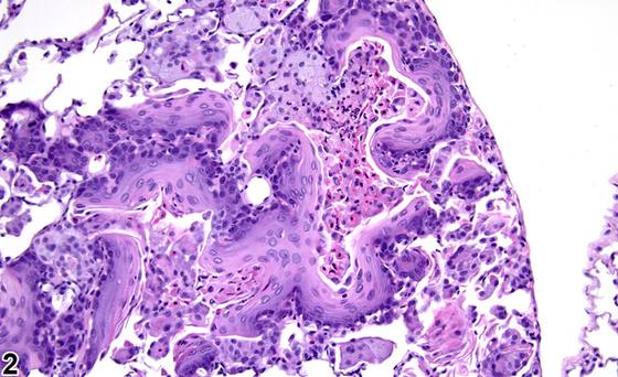
肺泡上皮的鳞状化生(squamous metaplasia)(Figure 1、Figure 2、Figure 3、Figure 4、Figure 5、Figure 6、Figure 7、Figure 8)以I型和II型肺泡上皮细胞(type I and type II pneumocytes)被鳞状上皮取代为特征。气道黏膜上皮的鳞状化生以细支气管或支气管上皮被鳞状上皮取代为特征。鳞状上皮是一种复层上皮,细胞从基底膜处的立方状基底细胞到表面的扁平鳞状细胞呈有序的成熟过程。上皮可发生角化,细胞可含有透明角质颗粒(keratohyalin granules)。早期鳞状化生仅表面细胞变扁平,可能难以识别。鳞状化生不应与再生混淆,因为再生的上皮细胞可呈扁平形态(伸展以覆盖裸露的基底膜)。通常,鳞状化生中存在一层以上的细胞。鳞状化生可见于对慢性刺激或损伤的反应,并被认为是一种适应性改变,因为鳞状上皮比呼吸道上皮或肺泡上皮对损伤更具抵抗力。因此,鳞状化生常伴有炎症、纤维化、坏死、溃疡以及上皮增生区域。虽然通常不被认为是肿瘤前病变,但在某些条件下鳞状化生可导致肿瘤形成(囊性角化上皮瘤或鳞状细胞癌)。存在非典型细胞的化生上皮(Figure 8)可能是肿瘤前病变。
诊断与分级建议(NTP 共识)
肺——化生,鳞状:只要存在即应诊断并赋予严重程度分级。诊断中应包含部位修饰词(即肺泡、细支气管或支气管)以指示病变的位置。相关病变,如炎症、纤维化、坏死或增生,应单独诊断。如果存在非典型性,则应在诊断中加入修饰词“非典型”(非典型性(atypical))。
图谱
英文原文
展开 NTP 官方英文描述与建议
Comment:
Squamous metaplasia of the alveolar epithelium ( Figure 1 , Figure 2 , Figure 3 , Figure 4 , Figure 5 , Figure 6 , Figure 7 , and Figure 8 ) is characterized by replacement of type I and type II pneumocytes with squamous epithelium. Squamous metaplasia of the airway epithelium is characterized by replacement of bronchiolar or bronchial epithelium with squamous epithelium. Squamous epithelium is a stratified epithelium with orderly progression of cell maturation from cuboidal basal cells at the basement membrane to flattened squamous cells at the surface. The epithelium may be keratinized, and the cells may contain keratohyalin granules. Early squamous metaplasia, where only the surface cells are flattened, can be difficult to identify. Squamous metaplasia should not be confused with regeneration, since regenerating epithelial cells can assume a flattened morphology (stretching out to cover a denuded basement membrane). Generally, more than one layer of cells is present in squamous metaplasia. Squamous metaplasia can be seen as a response to chronic irritation or injury and is thought to be an adaptive change, as the squamous epithelium is more resistant to injury than is respiratory or alveolar epithelium. Consequently, squamous metaplasia is often accompanied by inflammation, fibrosis, necrosis, ulceration, and areas of epithelial hyperplasia. Though not typically considered preneoplastic, under some conditions squamous metaplasia may give rise to neoplasia (cystic keratinizing epithelioma or squamous cell carcinoma). Metaplastic epithelium in which there are atypical cells ( Figure 8 ) may be preneoplastic.
Recommendation:
Lung - Metaplasia, Squamous should be diagnosed whenever present and assigned a severity grade. A site modifier (i.e., alveolus, bronchiole, or bronchus) should be included in the diagnosis to indicate the location of the lesion. Associated lesions, such as inflammation, fibrosis, necrosis, or hyperplasia, should be diagnosed separately. If atypia is present, then the modifier "atypical" should be added to the diagnosis.
来源与引用
原始条目:https://ntp.niehs.nih.gov/atlas/nnl/respiratory-system/lung/MetaplasiaSquamous
图谱总目录:NTP Nonneoplastic Lesion Atlas。 内容属美国联邦政府作品(公有领域),可引用;引用时请标注来源。
建议引用格式:NTP Nonneoplastic Lesion Atlas. Metaplasia Squamous (lung). LabSmart 中文版:
https://www.labsmart.cn/ntp/lung-metaplasia-squamous.html
中文图谱总页(可展开对照全部 595 条):https://www.labsmart.cn/ntp-atlas.html#lung-metaplasia-squamous