色素Pigment · 肺 · 呼吸系统 · NTP ntp-0116
诊断要点:当认为肺——色素与研究相关(或不确定是否与研究相关)或异常显著时,应予以诊断。并非所有色素都必须诊断,因为有些色素在老龄动物中普遍存在,或继发于其他疾病过程(如出血)而无毒理学意义。
病变描述
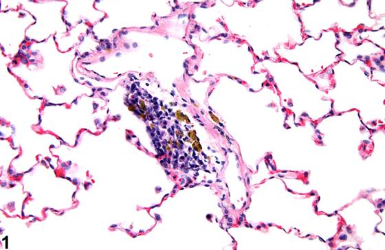
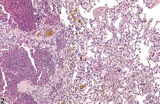
“色素(pigment)”一词专用于内源性物质。肺内可见多种类型的色素,包括含铁血黄素(hemosiderin)(Figure 1、Figure 2)和脂褐素(lipofuscin)。在肺内表现为棕色至黑色物质的受试物应诊断为异物(foreign material)而非色素(见肺——异物)。含铁血黄素是肺内最常见且最重要的色素类型。含铁血黄素在巨噬细胞内表现为细颗粒状、金棕色物质。当肺泡巨噬细胞吞噬红细胞时(如出血时发生),血红蛋白转化为含铁血黄素,即铁最稳定的储存形式。含铁血黄素见于出血之后,出血可由肺血管充血(如充血性心力衰竭时发生)或血管损伤引起。酸性血褐素(acid hematin)是低pH福尔马林固定产生的人工假象,表现为黑色至棕色无定形至微晶颗粒,易与含铁血黄素混淆。酸性血褐素由酸作用于血红蛋白产生,故可出现在出血区域。含铁血黄素的存在可通过Perls’普鲁士蓝染色技术确认。
诊断与分级建议(NTP 共识)
当认为肺——色素与研究相关(或不确定是否与研究相关)或异常显著时,应予以诊断。并非所有色素都必须诊断,因为有些色素在老龄动物中普遍存在,或继发于其他疾病过程(如出血)而无毒理学意义。病理学家应运用其判断决定是否应诊断该色素。当诊断色素时,应给出严重程度分级,并在适当时使用部位修饰词(如支气管、细支气管)以确定病变位置。即使使用一系列特殊染色,在组织学切片中确定色素鉴定通常也很困难。因此,诊断色素(而非诊断色素类型,如含铁血黄素或脂褐素)最为恰当。病理学描述应包括色素的形态学特征、其在组织内的分布以及受累细胞类型。相关病变,如出血、肺泡巨噬细胞浸润或炎症,应单独诊断。相关链接:肺——异物
图谱
英文原文
展开 NTP 官方英文描述与建议
Comment:
The term "pigment" is reserved for endogenous substances. Various types of pigment can be seen in the lungs, including hemosiderin ( Figure 1 and Figure 2 ) and lipofuscin. Test agents that appear as brown to black material in the lungs should be diagnosed as foreign material rather than pigment (see Lung - Foreign Material ). Hemosiderin is the most common and most important type of pigment seen in the lungs. Hemosiderin appears as finely granular, golden brown material in macrophages. When alveolar macrophages ingest erythrocytes, as occurs with hemorrhage, the hemoglobin is converted into hemosiderin, the most stable storage form of iron. Hemosiderin is seen following hemorrhage, which can result from vascular congestion in the lung (as occurs, e.g., with congestive heart failure) or vascular injury. Acid hematin, an artifact of fixation in formalin with a low pH, appears as black to brown amorphous to microcrystalline granules and can easily be confused with hemosiderin. Acid hematin is produced by acid acting on hemoglobin, so it may appear in areas of hemorrhage. The presence of hemosiderin can be confirmed with the Perls’ Prussian blue staining technique.
Recommendation:
Lung - Pigment should be diagnosed when it is considered to be relevant to the study (or if it is unclear whether it is relevant to the study) or if it is unusually prominent. Not all pigments have to be diagnosed, as some are ubiquitous in aging animals or are secondary to another disease process (e.g., hemorrhage) and not toxicologically meaningful. The pathologist should use his or her judgment in deciding whether or not the pigment should be diagnosed. When pigment is diagnosed, it should be given a severity grade, and if appropriate, a site modifier (e.g., bronchus, bronchiole) should be used to identify the location of the lesion. Definitive pigment identification is often difficult in histologic sections, even with a battery of special stains. Therefore, a diagnosis of pigment (as opposed to diagnosing the type of pigment, e.g., hemosiderin or lipofuscin) is most appropriate. The pathology narrative should include a description of the morphologic features of the pigment, its distribution within the tissue, and the cell type affected. Associated lesions, such as hemorrhage, alveolar macrophage infiltration, or inflammation, should be diagnosed separately. Related Links: Lung - Foreign Material
来源与引用
原始条目:https://ntp.niehs.nih.gov/atlas/nnl/respiratory-system/lung/Pigment
图谱总目录:NTP Nonneoplastic Lesion Atlas。 内容属美国联邦政府作品(公有领域),可引用;引用时请标注来源。
建议引用格式:NTP Nonneoplastic Lesion Atlas. Pigment (lung). LabSmart 中文版:
https://www.labsmart.cn/ntp/lung-pigment.html
中文图谱总页(可展开对照全部 595 条):https://www.labsmart.cn/ntp-atlas.html#lung-pigment