蛋白沉积症Proteinosis · 肺 · 呼吸系统 · NTP ntp-0117
诊断要点:Lung - Proteinosis 只要存在就应诊断,并根据病变范围进行分级。
病变描述
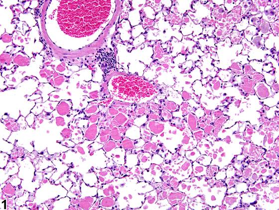
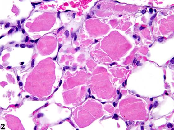
肺泡蛋白沉积症(alveolar proteinosis,Figure 1、Figure 2、Figure 3、Figure 4)的特征是肺泡内游离存在亮嗜酸性、无定形、periodic acid-Schiff 阳性的物质。通常,该物质伴随的炎症很少,但巨噬细胞数量可能增多。分布可为 局灶性(focal),仅少数肺泡受累;可为 patchy,累及肺叶的部分区域;或为 弥漫性(diffuse),累及整个肺叶。该物质被认为由脂质和肺蛋白组成。该病变的发病机制尚不清楚,但可能由表面活性物质产生增加和/或因巨噬细胞功能受损导致清除减少所致。肺泡蛋白沉积症必须与水肿液鉴别,水肿液更均质,在 hematoxylin and eosin 染色下嗜酸性染色不如前者明亮或深染,且 periodic acid-Schiff 阴性。肺泡蛋白沉积症可见于暴露于各种粉尘颗粒(如石英)的情况下。该病变在 rats 中似乎比在 mice 中更常见。
诊断与分级建议(NTP 共识)
Lung - Proteinosis 只要存在就应诊断,并根据病变范围进行分级。由于肺泡巨噬细胞常与该病变相关而增多,无需单独诊断,除非它们构成病变的显著成分。炎症应单独诊断,因为它通常与该病变无关。继发于肿瘤的蛋白沉积症不应诊断,但可在病理学叙述中描述。
图谱
英文原文
展开 NTP 官方英文描述与建议
Comment:
Alveolar proteinosis ( Figure 1 , Figure 2 , Figure 3 , and Figure 4 ) is characterized by brightly eosinophilic, amorphous, periodic acid-Schiff–positive material that is free within the alveoli. Typically, little inflammation is associated with the material, but there may be increased numbers of macrophages. The distribution may be focal, with few alveoli affected; patchy, affecting portions of lung lobes; or diffuse, affecting entire lobes. The material is thought to comprise lipids and pulmonary proteins. The pathogenesis of the lesion is not known but may be caused by increased surfactant production and/or decreased clearance due to macrophage impairment. Alveolar proteinosis must be distinguished from edema fluid, which is more homogeneous, does not stain as brightly or deeply eosinophilic with hematoxylin and eosin, and is periodic acid-Schiff negative. Alveolar proteinosis may be seen with exposure to various dust particles (e.g., quartz). The lesion appears to be more common in rats than in mice.
Recommendation:
Lung - Proteinosis should be diagnosed whenever present and graded based on the extent of the lesion. Since alveolar macrophages are often increased in association with this lesion, they need not be diagnosed separately unless they comprise a significant component of the lesion. Inflammation should be diagnosed separately since it is not typically associated with this lesion. Proteinosis that is secondary to a neoplasm should not be diagnosed but may be described in the pathology narrative.
来源与引用
原始条目:https://ntp.niehs.nih.gov/atlas/nnl/respiratory-system/lung/Proteinosis
图谱总目录:NTP Nonneoplastic Lesion Atlas。 内容属美国联邦政府作品(公有领域),可引用;引用时请标注来源。
建议引用格式:NTP Nonneoplastic Lesion Atlas. Proteinosis (lung). LabSmart 中文版:
https://www.labsmart.cn/ntp/lung-proteinosis.html
中文图谱总页(可展开对照全部 595 条):https://www.labsmart.cn/ntp-atlas.html#lung-proteinosis