溃疡形成Ulceration · 肺 · 呼吸系统 · NTP ntp-0105
诊断要点:肺 – 溃疡:只要存在即应诊断并赋予严重程度分级。诊断中应包含部位修饰词(例如支气管、细支气管、肺泡),以指示病变的位置。
病变描述
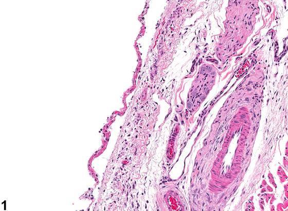
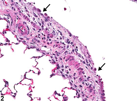
支气管或细支气管上皮的溃疡(ulceration)表现为被覆上皮的缺失(Figure 1、Figure 2),暴露出下方的基底膜。必须区分溃疡与坏死,以及溃疡与再生。如果坏死细胞仍附着于基底膜,则应视为坏死而非溃疡。如果坏死细胞已被存活的上皮细胞所取代,这些细胞伸展并覆盖受累区域,则应视为再生。溃疡通常由上皮细胞损伤引起,损伤原因可以是直接毒性,也可以与炎症相关,在炎症中上皮细胞损伤可能由活性氧或其他介质所致。尽管急性溃疡可能不伴炎症,但溃疡通常伴有混合性炎症反应。在呼吸道上皮坏死和溃疡的部位,常可见纤维素性渗出物。在某些病例中,成纤维细胞可浸润渗出物,导致纤维化,严重病例中纤维化可跨越管腔(管腔内纤维化,intraluminal fibrosis)并阻塞气流。仙台病毒感染(Sendai virus infection)可引起支气管和细支气管上皮的溃疡。
诊断与分级建议(NTP 共识)
肺 – 溃疡:只要存在即应诊断并赋予严重程度分级。诊断中应包含部位修饰词(例如支气管、细支气管、肺泡),以指示病变的位置。如果坏死细胞仍附着于气道表面,则应诊断坏死;如果相邻细胞已变平并迁移覆盖溃疡面,则应诊断再生(参见 Lung - Epithelium - Necrosis 和 Lung - Regeneration)。相关病变,如炎症,除非严重程度确有必要,否则不应单独诊断,但应在病理学叙述中描述。相关链接:Lung - Epithelium - Necrosis Lung - Regeneration
图谱
英文原文
展开 NTP 官方英文描述与建议
Comment:
Ulceration of the bronchial or bronchiolar epithelium represents loss of the lining epithelium ( Figure 1 and Figure 2 ) exposing the underlying basement membrane. A distinction must be made between ulceration and necrosis and between ulceration and regeneration. If necrotic cells remain attached to the basement membrane, this would be considered necrosis rather than an ulcer. If the necrotic cells have been replaced by viable epithelial cells that have stretched across and cover the affected region, this would be considered regeneration. Ulceration is typically caused by damage to the epithelial cells either by direct toxicity or in association with inflammation in which epithelial cell damage may be caused by reactive oxygen species or other mediators. Though there may be no inflammation with acute ulcers, ulcers are typically associated with a mixed inflammatory response. A fibrinous exudate can often be seen at the site of respiratory epithelial necrosis and ulceration. In some cases, fibroblasts can infiltrate the exudate, resulting in fibrosis, which in severe cases can cross the lumen (intraluminal fibrosis) and obstruct air flow. Sendai virus infection can cause ulceration of bronchial and bronchiolar epithelium.
Recommendation:
Lung – Ulceration should be diagnosed and assigned a severity grade whenever present. A site modifier (e.g., bronchus, bronchiole, alveolus) should be included in the diagnosis to indicate the location of the lesion. If the necrotic cells are still attached to the airway surface, then necrosis should be diagnosed; if the adjacent cells have flattened and migrated across the ulcer, then regeneration should be diagnosed (see Lung - Epithelium - Necrosis and Lung - Regeneration ). Associated lesions, such as inflammation, should not be diagnosed separately unless warranted by severity but should be described in the pathology narrative. Related Links: Lung - Epithelium - Necrosis Lung - Regeneration
来源与引用
原始条目:https://ntp.niehs.nih.gov/atlas/nnl/respiratory-system/lung/Ulceration
图谱总目录:NTP Nonneoplastic Lesion Atlas。 内容属美国联邦政府作品(公有领域),可引用;引用时请标注来源。
建议引用格式:NTP Nonneoplastic Lesion Atlas. Ulceration (lung). LabSmart 中文版:
https://www.labsmart.cn/ntp/lung-ulceration.html
中文图谱总页(可展开对照全部 595 条):https://www.labsmart.cn/ntp-atlas.html#lung-ulceration