鳞状增生Hyperplasia Squamous · 口腔黏膜 · 消化系统 · NTP ntp-0228
诊断要点:鳞状细胞增生应当被诊断,并根据增生区域的大小、厚度和数量进行分级。病变的位置(例如牙龈、腭部)应在病理描述中注明。
病变描述
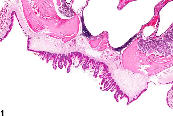
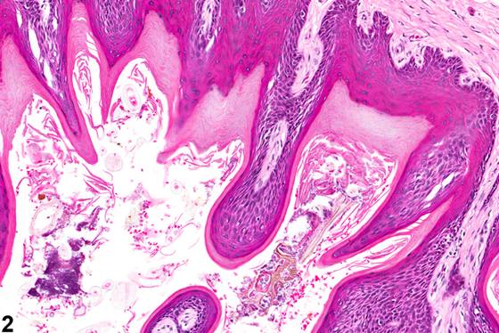
口腔黏膜的鳞状细胞增生(squamous hyperplasia)通常见于腭部(Figure 1、Figure 2、Figure 3)或牙龈(Figure 4)。鳞状细胞增生的特征是鳞状细胞数量增多,导致鳞状上皮厚度增加,可呈弥漫性(diffuse)或斑块状,或形成钝的乳头状突起。角化过度(hyperkeratosis)常与鳞状细胞增生同时出现。这些病变可能发生溃疡,通常归因于机械性摩擦。鳞状上皮的增生性病变被认为是一个连续的过程,从局灶性(focal)增生进展为乳头状瘤(papilloma),再进展为浸润性鳞状细胞癌(squamous cell carcinoma),尽管乳头状瘤阶段可能被跳过,直接从局灶性增生进展为鳞状细胞癌。乳头状瘤是表浅肿瘤,具有显著(marked)棘层肥厚(acanthosis)、角化过度和乳头状结构。乳头状瘤通常呈外生性(exophytic),但内翻性乳头状瘤(inverted papilloma)呈杯状向下方间质内生长。外生性乳头状瘤具有由结缔组织构成的血管化核心,从黏膜下层延伸(形成蒂样结构),表面覆盖增生的鳞状上皮细胞,这些细胞常发生角化。无蒂形态(sessile shape)、细胞分化良好的外观以及缺乏显著的间质成分,是区分增生与乳头状瘤的重要依据。鳞状细胞癌与增生的区别在于,鳞状细胞癌的肿瘤细胞通常为异型细胞,伴有核增大(karyomegaly)、核极性丧失、核仁增大显著以及核分裂象增多。肿瘤性鳞状细胞突破基底膜浸润,肿瘤性上皮细胞以岛状、索状和梁状延伸至下方间质。
诊断与分级建议(NTP 共识)
鳞状细胞增生应当被诊断,并根据增生区域的大小、厚度和数量进行分级。病变的位置(例如牙龈、腭部)应在病理描述中注明。相关病变,如炎症,除非严重程度需要,否则不应单独诊断。与增生相关的角化过度或溃疡不应单独诊断,而应作为病理描述中病变描述的一部分。
图谱
英文原文
展开 NTP 官方英文描述与建议
Comment:
Squamous hyperplasia of the oral mucosa is usually seen on the palate ( Figure 1 , Figure 2 , and Figure 3 ) or gingiva ( Figure 4 ). Squamous hyperplasia is characterized by increased numbers of squamous cells resulting in increased thickness of the squamous epithelium, which may be diffuse or plaque-like or form blunt papillary projections. Hyperkeratosis is frequently seen with squamous cell hyperplasia. Ulceration of the these lesions may be seen and is usually attributed to mechanical abrasion. Proliferative lesions of the squamous epithelium are thought to be on a continuum, progressing from focal hyperplasia to papilloma to invasive squamous cell carcinoma, although the papilloma stage may be skipped, with progress directly from focal hyperplasia to squamous cell carcinoma. Papillomas are superficial tumors with marked acanthosis, hyperkeratosis, and papillary patterns. Papillomas are usually exophytic, although the inverted papilloma grows in a cuplike shape into the underlying stroma. Exophytic papillomas have a vascularized inner core of connective tissue extending from the submucosa (forming a stalklike structure) that is covered by proliferating squamous epithelial cells, which are often keratinized. The sessile shape, well-differentiated appearance of cells, and absence of a prominent stromal component are important in distinguishing hyperplasia from papilloma. Squamous cell carcinomas can be differentiated from hyperplasia in that the neoplastic cells of a squamous cell carcinoma generally have atypical cells with karyomegaly, loss of nuclear polarity, enlarged prominent nucleoli, and increased mitotic figures. Neoplastic squamous cells invade beyond the basement membrane, with islands, cords, and trabeculae of neoplastic epithelial cells extending into the underlying stroma.
Recommendation:
Squamous hyperplasia should be diagnosed and graded based on the size, thickness, and number of hyperplastic areas. The location of the lesion (e.g., gingival, palatal) should be noted in the pathology narrative. Associated lesions, such as inflammation, should not be diagnosed separately unless warranted by severity. Hyperkeratosis or ulceration associated with hyperplasia should not be diagnosed separately but should be a part of the lesion description in the pathology narrative.
来源与引用
原始条目:https://ntp.niehs.nih.gov/atlas/nnl/alimentary-system/oral-mucosa/HyperplasiaSquamous
图谱总目录:NTP Nonneoplastic Lesion Atlas。 内容属美国联邦政府作品(公有领域),可引用;引用时请标注来源。
建议引用格式:NTP Nonneoplastic Lesion Atlas. Hyperplasia Squamous (oral-mucosa). LabSmart 中文版:
https://www.labsmart.cn/ntp/oral-mucosa-hyperplasia-squamous.html
中文图谱总页(可展开对照全部 595 条):https://www.labsmart.cn/ntp-atlas.html#oral-mucosa-hyperplasia-squamous