增生Hyperplasia · 胰岛 · 内分泌系统 · NTP ntp-0092
诊断要点:胰岛细胞增生存在时应记录并给予严重程度分级。严重程度分级由受累胰岛的大小和数量共同确定。
病变描述
在小鼠中,胰岛大小差异很大,直径从50至300微米不等。由于小鼠的胰岛被认为是一个增殖群体,倍增时间为500天,因此其大小预期会随年龄增长而增加。由于这种大小差异,可能难以明确诊断增生。当胰岛更为显著时,可能看起来数量有所增加。然而,据报道小鼠每个胰腺中胰岛数量相对恒定,约为1,000个。胰岛细胞增生可自发发生于小鼠和大鼠,并且在小鼠中可能表现显著。它通常与年龄相关,并且通常累及多个胰岛。在大鼠中,胰岛细胞增生在雄性中比雌性更常见。如果有数个胰岛直径大于300微米,应作出胰岛细胞增生的诊断。此外,如果胰腺某切片中胰岛占据的总面积大于对照组,则作出胰岛细胞增生的诊断可能是恰当的。受累胰岛可能相互连续,呈小叶状外观(Figure 3),或明显分离(Figure 4)。增生与腺瘤之间的区别在很大程度上受腺瘤中存在压迫证据而增生中缺乏压迫的影响(Figure 5、Figure 6)。在胰岛细胞腺瘤中,可能存在一些细胞异型性或多形性,这在增生性病变中通常不见。偶见的腺泡细胞可能陷入融合的增生性胰岛中(Figure 7、Figure 8、Figure 9),不应与胰岛细胞癌的浸润相混淆。
诊断与分级建议(NTP 共识)
胰岛细胞增生存在时应记录并给予严重程度分级。严重程度分级由受累胰岛的大小和数量共同确定。如果在某一研究中存在治疗相关效应和/或胰岛腺瘤的证据,病理学描述应提供用于区分增生与腺瘤的诊断特征(例如,增生中缺乏压迫、异型性或坏死),并说明任何治疗相关反应的程度。
图谱
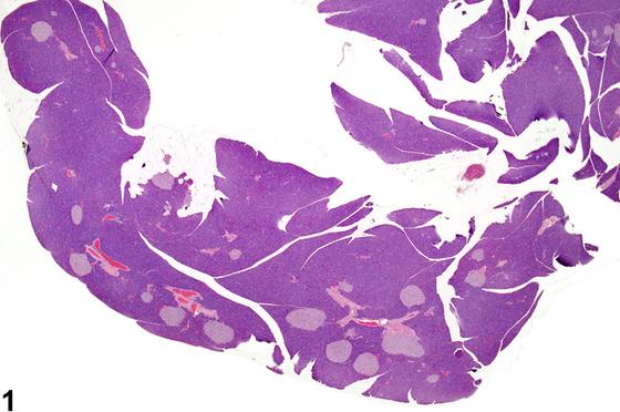
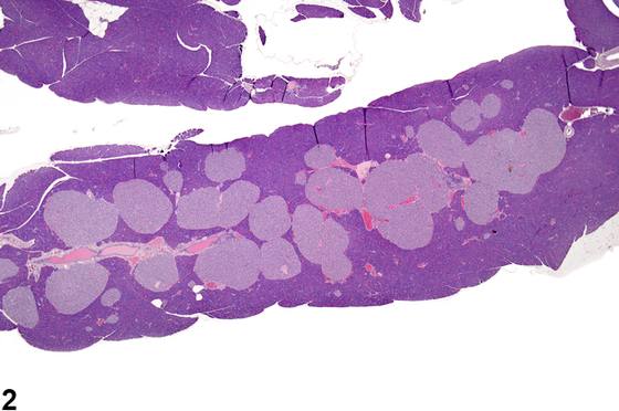
英文原文
展开 NTP 官方英文描述与建议
Comment:
In mice, islets vary greatly in size, ranging from 50 to 300 micrometers in diameter. Since islets in mice are considered to be an expanding population with a doubling time of 500 days, their size would be expected to increase with age. Because of this size variation, it may be difficult to definitively diagnose hyperplasia. When islets are more prominent, they may appear to have increased in number. However, mice are reported to have a relatively constant 1,000 islets per pancreas. Islet cell hyperplasia occurs spontaneously in mice and rats and may be dramatic in mice. It is often age associated and typically involves multiple islets. In rats, islet cell hyperplasia occurs more frequently in males than females. If there several islets greater than 300 micrometers in diameter, a diagnosis of islet cell hyperplasia should be made. Also, if the overall area occupied by the islets in a section of pancreas is greater than that of the controls, a diagnosis of islet cell hyperplasia may be appropriate. Affected islets may be contiguous, giving a lobular appearance ( Figure 3 ) or distinctly separated ( Figure 4 ). The distinction between hyperplasia and adenoma is largely influenced by evidence of compression in adenomas and lack of compression in hyperplasia ( Figure 5 and Figure 6 ). In islet cell adenomas, there may be some cellular atypia or pleomorphism, which is not typically seen in hyperplastic lesions. Occasional acinar cells may become trapped within coalescing hyperplastic islets ( Figure 7 , Figure 8 , and Figure 9 ) and should not be confused with invasion of an islet cell carcinoma.
Recommendation:
Islet cell hyperplasia should be documented when present and given a severity grade. Severity grading is determined by both size and number of affected islets. If there is evidence of treatment-related effect and/or the presence of islet adenomas in a given study, the pathology narrative should provide the diagnostic features used to distinguish between hyperplasia and adenoma (e.g., lack of compression, atypia, or necrosis in hyperplasia) and address the magnitude of any treatment-related response.
来源与引用
原始条目:https://ntp.niehs.nih.gov/atlas/nnl/endocrine-system/pancreatic-islets/Hyperplasia
图谱总目录:NTP Nonneoplastic Lesion Atlas。 内容属美国联邦政府作品(公有领域),可引用;引用时请标注来源。
建议引用格式:NTP Nonneoplastic Lesion Atlas. Hyperplasia (pancreatic-islets). LabSmart 中文版:
https://www.labsmart.cn/ntp/pancreatic-islets-hyperplasia.html
中文图谱总页(可展开对照全部 595 条):https://www.labsmart.cn/ntp-atlas.html#pancreatic-islets-hyperplasia