萎缩Atrophy · 唾液腺 · 消化系统 · NTP ntp-0247
诊断要点:无论何时出现,萎缩均应予以诊断,并根据导管/腺泡细胞萎缩的程度及唾液腺内受累范围进行分级。
病变描述
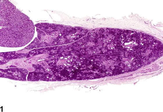
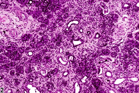
唾液腺萎缩(Figure 1、Figure 2)常见于老年F344/N大鼠。与许多其他腺体器官一样,分泌腺的大小对功能需求具有应答性,并受年龄相关变化的影响。在老龄大鼠中,颗粒(卷曲)管的数量和上皮高度以及成熟分泌颗粒的数量已被证明随年龄增长而减少。间质纤维结缔组织可能轻度增多,并可能有慢性炎性细胞浸润。有时在小叶和腺泡之间可见局灶性脂肪细胞聚集,类似于胰腺和胸腺的退行性变化。也可能存在少数单个腺泡细胞的坏死。其他品系大鼠中可见与F344大鼠相似的年龄相关萎缩性或退行性变化。萎缩可能是毒性的结果,但常见与唾液腺肿瘤或炎性病变相关。当细胞体积减小呈弥漫性时,萎缩可能很轻微且易被忽视。萎缩、坏死和炎症通常见于活动性涎泪腺炎病毒(sialodacryoadenitis virus)感染。萎缩也可见于单核细胞白血病浸润唾液腺或面神经分支起源的神经鞘瘤局部侵袭时。导管腔内物质的矿化不常发生,并可与萎缩同时出现。去神经支配、肾上腺素能拮抗剂、导管结扎或流质饮食可导致唾液腺萎缩。饮食因素影响唾液腺大小。食物摄入减少或蛋白质饥饿可降低大鼠唾液腺重量。黏液腺和浆液腺的皱缩及酶原颗粒的丢失与RNA减少但DNA含量不变相关,归因于蛋白质合成需求降低。由于唾液腺功能对肾上腺素能刺激具有应答性,因此肾上腺素能阻断后发生萎缩并不令人意外。小鼠颌下腺重量在给予β-肾上腺素能阻断剂普萘洛尔(propranolol)后显示下降。这与可染性中性黏蛋白减少及腺泡细胞厚度减小相关,使腺腔显得比正常更大。唾液腺萎缩偶尔发生于老龄B6C3F1小鼠。萎缩一般累及单个小叶,其特征为腺泡和腺泡上皮细胞体积减小,可伴有慢性炎性细胞浸润及间质结缔组织和/或腺管数量增加。颌下腺和腮腺比舌下腺更常受累。
诊断与分级建议(NTP 共识)
无论何时出现,萎缩均应予以诊断,并根据导管/腺泡细胞萎缩的程度及唾液腺内受累范围进行分级。与年龄相关萎缩有关的炎症和纤维化不单独诊断,除非其严重程度足以支持单独诊断。
图谱
英文原文
展开 NTP 官方英文描述与建议
Comment:
Atrophy of the salivary glands ( Figure 1 and Figure 2 ) occurs commonly in older F344/N rats. As is the case with many other glandular organs, the size of the secretory glands is responsive to functional demand and is subject to age-related changes. In aging rats, the number and epithelial height of granular (convoluted) ducts and the number of mature secretory granules have been shown to decrease with age. There may be a slight increase in stromal fibrous connective tissue, and there may be an infiltrate of chronic inflammatory cells. Focal aggregates of adipocytes are sometimes seen between lobules and acini, similar to involutional changes in the pancreas and thymus. Necrosis of a few individual acinar cells may also be present. Age-associated atrophic or degenerative changes similar to those in the F344 rat are seen in other strains of rats. Atrophy may be the result of toxicity, but it is commonly seen in association with neoplasia or inflammatory lesions in the salivary gland. Atrophy can be subtle and can be overlooked when the decrease in cell size is diffuse. Atrophy, necrosis, and inflammation are typically seen with active sialodacryoadenitis virus infection. Atrophy is also seen with mononuclear cell leukemia infiltration of the salivary gland or local invasion by a schwannoma arising in branches of the facial nerve. Mineralization of material in the ductular lumen occurs infrequently and may be seen with atrophy. Denervation, adrenergic antagonists, duct ligation, or liquid diets can result in salivary gland atrophy. Dietary factors influence salivary gland size. Decreased food consumption or protein starvation can reduce the weight of salivary glands in rats. Shrinking of mucous and serous glands and loss of zymogen granules are associated with decreased RNA but unchanged DNA content, attributable to the reduced requirements for protein synthesis. As salivary gland function is responsive to adrenergic stimulation, it is not surprising that atrophy occurs following adrenergic blockage. The weights of the submandibular gland in mice were shown to decrease after administration of the B-adrenergic blocking agent propranolol. This was associated with a reduction in stainable neutral mucins and a decrease in the thickness of the acinar cells, making the gland lumens appear larger than normal. Salivary gland atrophy occurs occasionally in aging B6C3F1 mice. The atrophy generally affects individual lobules and is characterized by a decrease in the size of acini and acinar epithelial cells, which may be accompanied by an infiltrate of chronic inflammatory cells and an increase in interstitial connective tissue and/or in the number of glandular ducts. The submandibular and parotid glands tend to be involved more commonly than the sublingual gland.
Recommendation:
Whenever present, atrophy should be diagnosed and graded based on the degree of ductal/acinar cell atrophy and the extent of involvement within the salivary gland. Inflammation and fibrosis associated with age related atrophy are not diagnosed separately unless warranted by severity.
来源与引用
原始条目:https://ntp.niehs.nih.gov/atlas/nnl/alimentary-system/salivary-gland/Atrophy
图谱总目录:NTP Nonneoplastic Lesion Atlas。 内容属美国联邦政府作品(公有领域),可引用;引用时请标注来源。
建议引用格式:NTP Nonneoplastic Lesion Atlas. Atrophy (salivary-gland). LabSmart 中文版:
https://www.labsmart.cn/ntp/salivary-gland-atrophy.html
中文图谱总页(可展开对照全部 595 条):https://www.labsmart.cn/ntp-atlas.html#salivary-gland-atrophy