细胞浸润Infiltration Cellular · 唾液腺 · 消化系统 · NTP ntp-0243
诊断要点:无论何时出现,唾液腺中的细胞浸润应诊断为记录为“infiltration, cellular”,并指明适当的细胞类型(即淋巴细胞、嗜酸性粒细胞、组织细胞等)。
病变描述
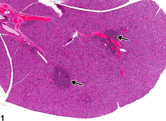
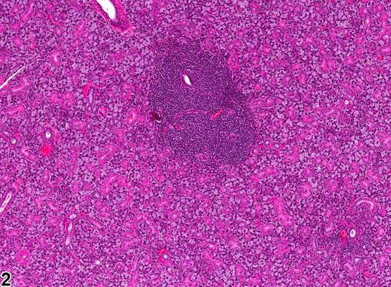
唾液腺中的浸润细胞可包括小型成熟淋巴细胞、嗜酸性粒细胞(eosinophil)、肥大细胞、组织细胞和未退变的粒细胞。单核细胞浸润,主要为淋巴细胞,有时混有少量浆细胞,在B6C3F1小鼠的唾液腺中常见(Figure 1、Figure 2),如同在B6C3F1小鼠的其他器官中一样。这些浸润在唾液腺中的意义尚不清楚。浸润可由单个或多个细胞簇组成,通常在雌性中较大且更频繁。它们一般始于血管周围间隙,但可能增大至浸润并取代腺实质。在发生年龄相关唾液腺萎缩的老年大鼠中,可能存在淋巴细胞或浆细胞灶。有必要将显著(marked)淋巴细胞浸润与慢性炎症和淋巴瘤区分开来。慢性炎症还会伴有坏死和组织损伤,而这些在细胞浸润中未见。淋巴瘤预期会导致唾液腺结构消失,并存在于其他器官,因为淋巴瘤罕见于唾液腺原发。
诊断与分级建议(NTP 共识)
无论何时出现,唾液腺中的细胞浸润应诊断为记录为“infiltration, cellular”,并指明适当的细胞类型(即淋巴细胞、嗜酸性粒细胞、组织细胞等)。严重程度分级基于浸润唾液腺的细胞数量。如果淋巴细胞(或其他浸润细胞)被认为是萎缩的组成部分,则不应诊断,而应在叙述中随萎缩一起描述。
图谱
英文原文
展开 NTP 官方英文描述与建议
Comment:
Infiltrates in the salivary gland can include small mature lymphocytes, eosinophils, mast cells, histiocytes, and nondegenerate granulocytes. Infiltrates of mononuclear cells, primarily lymphocytes, sometimes admixed with a few plasma cells, occur commonly in the salivary glands of B6C3F1 mice ( Figure 1 and Figure 2 ), as in other organs in the B6C3F1 mouse. The significance of these infiltrates in the salivary gland is unclear. The infiltrates may consist of a single or multiple clusters and are usually larger and more frequent in females. They generally begin in the perivascular space but may increase in size so as to infiltrate and displace glandular parenchyma. Foci of lymphocytes or plasma cells may be present in older rats undergoing age-related atrophy of the salivary glands. It is necessary to differentiate marked lymphocyte infiltrate from chronic inflammation and lymphoma. Chronic inflammation will also have necrosis and tissue damage, which are not noted with cellular infiltrates. Lymphoma would be expected to result in effacement of salivary gland architecture and to be present in other organs, as lymphoma is rarely primary to the salivary gland.
Recommendation:
Whenever present, an infiltrate of cells in the salivary gland should be diagnosed as recorded as "infiltration, cellular" with the appropriate cell type indicated (i.e., lymphocyte, eosinophil, histiocyte, etc.). Severity grading is based on the number of cells infiltrating the salivary gland. If lymphocytes (or other infiltrating cells) are considered a component of atrophy, they should not be diagnosed but should be described in the narrative with the atrophy.
来源与引用
原始条目:https://ntp.niehs.nih.gov/atlas/nnl/alimentary-system/salivary-gland/InfiltrationCellular
图谱总目录:NTP Nonneoplastic Lesion Atlas。 内容属美国联邦政府作品(公有领域),可引用;引用时请标注来源。
建议引用格式:NTP Nonneoplastic Lesion Atlas. Infiltration Cellular (salivary-gland). LabSmart 中文版:
https://www.labsmart.cn/ntp/salivary-gland-infiltration-cellular.html
中文图谱总页(可展开对照全部 595 条):https://www.labsmart.cn/ntp-atlas.html#salivary-gland-infiltration-cellular