纤维化Fibrosis · 皮肤 · 皮肤系统 · NTP ntp-0373
诊断要点:在大多数病例中,纤维化应当被记录并赋予严重程度分级。作为慢性或慢性活动性炎症的次要成分而存在的纤维化,或被认为是继发于表皮溃疡的纤维化,可能不必单独诊断,但应在病理学叙述中予以描述。
病变描述
纤维化通常是因慢性化学暴露或创伤所致表皮或真皮损伤的后遗症。其特征为真皮(Figure 1)或皮下组织中纤维性结缔组织增多,并且通常伴有慢性炎症。纤维化可能表现为一种细微的病变,尤其是在后期阶段。其特征为真皮内或毛囊周围成纤维细胞和胶原纤维增生,通常与表皮平行排列。在更严重的病例中,纤维化可向真皮和皮下组织深部延伸。与无纤维化的真皮相比,纤维化区域可能呈现轻度嗜碱性外观,但这可因染色质量和纤维化病变的成熟程度而异。在早期纤维化中,成纤维细胞通常伴有炎性细胞(尤其是当与表皮溃疡相关时);成纤维细胞往往较大且活性较高,胶原纤维较不致密且排列更紊乱。在纤维化的后期阶段,成纤维细胞往往较小且更呈梭形;胶原纤维排列更有组织且更致密,并且炎症通常较少。在严重病例中,纤维化区域可能出现毛囊缺失。
诊断与分级建议(NTP 共识)
在大多数病例中,纤维化应当被记录并赋予严重程度分级。作为慢性或慢性活动性炎症的次要成分而存在的纤维化,或被认为是继发于表皮溃疡的纤维化,可能不必单独诊断,但应在病理学叙述中予以描述。当炎症程度与纤维化程度不相称时(如Figure 1所示),则应当对纤维化进行诊断和分级。可能与纤维化同时见到的其他相关病变,包括表皮增生,应当单独诊断。
图谱
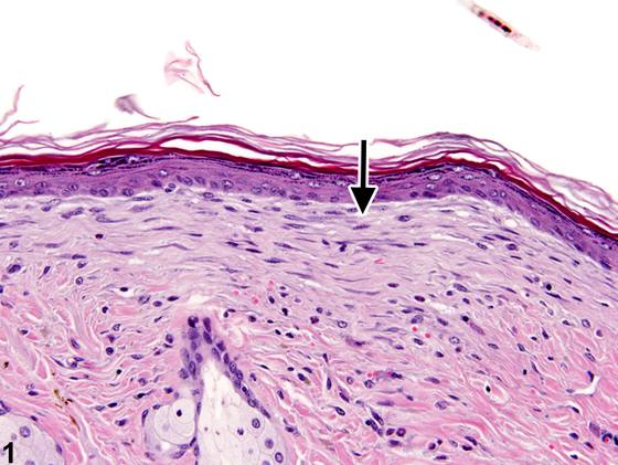
英文原文
展开 NTP 官方英文描述与建议
Comment:
Fibrosis is often a sequela of epidermal or dermal injury due to chronic chemical exposure or trauma. It is characterized by an increase of fibrous connective tissues in the dermis ( Figure 1 ) or subcutis and usually accompanies chronic inflammation. Fibrosis may occur as a subtle finding, particularly in the later stages. It is characterized by proliferation of fibroblasts and collagen fibers in the dermis or around hair follicles, typically oriented parallel to the epidermis. In more severe cases, the fibrosis can extend deeper into the dermis and subcutis. Areas of fibrosis may have a slightly basophilic appearance compared with the nonfibrotic dermis, but this can vary with staining quality and the maturity of the fibrotic lesion. In early fibrosis, the fibroblasts are usually accompanied by inflammatory cells (particularly when associated with an epidermal ulcer); the fibroblasts tend to be larger and more active, and the collagen fibers are less compact and more disorganized. In the later stages of fibrosis, the fibroblasts tend to be smaller and more spindle shaped; the collagen fibers are more organized and compact, and there is often less inflammation. In severe cases, there may be a loss of hair follicles in the fibrotic region.
Recommendation:
Fibrosis should be documented and assigned a severity grade in most cases. Fibrosis that is present as a minor component of chronic or chronic active inflammation or is considered secondary to epidermal ulceration may not warrant a separate diagnosis but should be described in the pathology narrative. When the degree of inflammation is not commensurate to that of fibrosis (as in Figure 1 ), then fibrosis should be diagnosed and graded. Other associated lesions that may be seen with fibrosis, including epidermal hyperplasia, should be diagnosed separately.
来源与引用
原始条目:https://ntp.niehs.nih.gov/atlas/nnl/integumentary-system/skin/Fibrosis
图谱总目录:NTP Nonneoplastic Lesion Atlas。 内容属美国联邦政府作品(公有领域),可引用;引用时请标注来源。
建议引用格式:NTP Nonneoplastic Lesion Atlas. Fibrosis (skin). LabSmart 中文版:
https://www.labsmart.cn/ntp/skin-fibrosis.html
中文图谱总页(可展开对照全部 595 条):https://www.labsmart.cn/ntp-atlas.html#skin-fibrosis