增生Hyperplasia · 皮肤 · 皮肤系统 · NTP ntp-0367
诊断要点:首选“上皮增生(epithelial hyperplasia)”而非“棘层肥厚(acanthosis)”一词。
病变描述
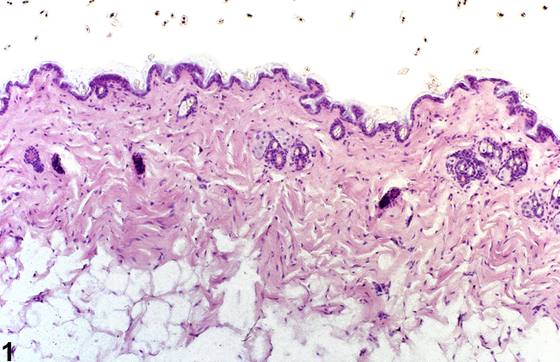
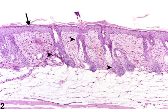
上皮增生(epithelial hyperplasia)是NTP研究中在B6C3F1小鼠皮肤观察到的最常见的自发性非肿瘤性病变。表皮及附属器上皮的增生也是皮肤局部涂抹化学物质后的常见反应。在较严重的病例中,尤其是伴有炎症时,常发生毛囊上皮、毛囊单位及皮脂腺的增生。其特征为:与对照动物的相同部位(Figure 1)相比,表皮非角化层因上皮细胞数量(层数)增加而增厚(Figure 2)。毛囊上皮也可受累。这种增厚可累及全部三个非角化层(基底层、棘层和颗粒层),但可能在棘细胞层最为明显。在较严重的病例中,毛囊上皮也可发生增生。该病变可继发于多种损伤和状况,包括自发性和实验诱发的炎症过程、暴露于刺激物或毒性物质、浅表角化层的反复磨损以及长期暴露于紫外线。皮脂腺增生的特征为皮脂腺增大,细胞数量增加,在中央导管周围形成众多小叶(Figure 3、Figure 4)。在为期2年的研究期间,皮脂腺增生有可能进展为良性和恶性皮脂腺细胞肿瘤。毛囊增生的特征为真皮和皮下组织内毛囊单位数量增加(Figure 5)。毛囊上皮增生的特征为毛囊内毛囊上皮细胞数量增加;它通常是表皮上皮增生的延伸,但也可见于无表皮增生的情况下。
诊断与分级建议(NTP 共识)
首选“上皮增生(epithelial hyperplasia)”而非“棘层肥厚(acanthosis)”一词。只要存在,上皮增生即应作出诊断并赋予严重程度分级。当毛囊上皮也发生增生时,这一点应反映在赋予上皮增生的严重程度分级中,并在病理学描述中加以描述。如果毛囊上皮增生发生而无表皮受累,则应使用“皮肤,毛囊,上皮——增生(Skin, Hair follicle, Epithelium – Hyperplasia)”一词。相关病变,如炎症,应单独诊断。当作为再生性反应、继发于邻近溃疡而存在时,增生不应作出诊断,但应在描述中加以描述。当存在时,皮脂腺增生或毛囊增生应作出诊断并赋予严重程度分级。皮脂腺细胞的增生常伴有肥大。在此类情况下,肥大无需诊断。
图谱
英文原文
展开 NTP 官方英文描述与建议
Comment:
Epithelial hyperplasia is the most common spontaneous, non-neoplastic lesion of the skin observed in B6C3F1 mice in NTP studies. Hyperplasia of the epithelium of the epidermis and adnexa is also a common response to dermal application of chemicals. In more severe cases, especially when accompanied by inflammation, hyperplasia of follicular epithelium, follicular units, and sebaceous glands often occurs. It is characterized by increased thickness of the nonkeratinized layers of the epidermis due to an increased number (layers) of epithelial cells ( Figure 2 ) compared with the same site in a control animal ( Figure 1 ). The epithelium of the hair follicles may also be affected. This increased thickness may involve all three nonkeratinized layers (basal, spinous, and granular) but may be most apparent in the spinous cell layer. In more severe cases, the follicular epithelium may also be hyperplastic ( Figure 2 ). This lesion can result after a wide variety of injuries and conditions, including spontaneous and experimentally induced inflammatory processes, exposure to irritants or toxic materials, repeated abrasion of the superficial keratin layer, and prolonged exposure to ultraviolet light. Sebaceous gland hyperplasia is characterized by large sebaceous glands with increased numbers of cells forming numerous lobules around central ducts ( Figure 3 and Figure 4 ). Over the period of a 2-year study, sebaceous hyperplasia has the potential to progress to benign and malignant sebaceous cell neoplasms. Follicular hyperplasia is characterized by increased numbers of follicular units within the dermis and subcutis ( Figure 5 ). Follicular epithelial hyperplasia is characterized by increased numbers of follicular epithelial cells within the hair follicles; it is typically an extension of epithelial hyperplasia of the epidermis but may be seen in the absence of epidermal hyperplasia.
Recommendation:
“Epithelial hyperplasia” is preferred over the term “acanthosis.” Whenever present, epithelial hyperplasia should be diagnosed and assigned a severity grade. When follicular epithelium is also hyperplastic, this should be reflected in the severity grade assigned to epithelial hyperplasia and described in the pathology narrative. If follicular epithelial hyperplasia occurs without epidermal involvement, the term “Skin, Hair follicle, Epithelium – Hyperplasia” should be used. Associated lesions, such as inflammation, should be diagnosed separately. When present as a regenerative response, secondary to an adjacent ulcer, hyperplasia should not be diagnosed but should be described in the narrative. When present, sebaceous hyperplasia or follicular hyperplasia should be diagnosed and assigned a severity grade. Hyperplasia of the sebaceous cells is often accompanied by hypertrophy. In such cases, hypertrophy need not be diagnosed.
来源与引用
原始条目:https://ntp.niehs.nih.gov/atlas/nnl/integumentary-system/skin/Hyperplasia
图谱总目录:NTP Nonneoplastic Lesion Atlas。 内容属美国联邦政府作品(公有领域),可引用;引用时请标注来源。
建议引用格式:NTP Nonneoplastic Lesion Atlas. Hyperplasia (skin). LabSmart 中文版:
https://www.labsmart.cn/ntp/skin-hyperplasia.html
中文图谱总页(可展开对照全部 595 条):https://www.labsmart.cn/ntp-atlas.html#skin-hyperplasia