坏死Necrosis · 皮肤 · 皮肤系统 · NTP ntp-0366
诊断要点:观察到上皮坏死时,应作出诊断并赋予严重程度分级。在较严重病例中,毛囊上皮也可发生坏死;
病变描述
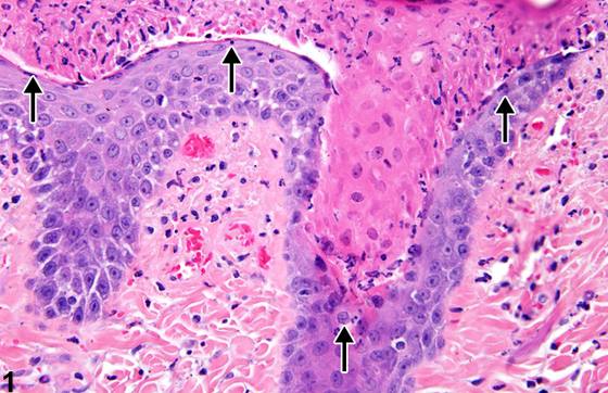
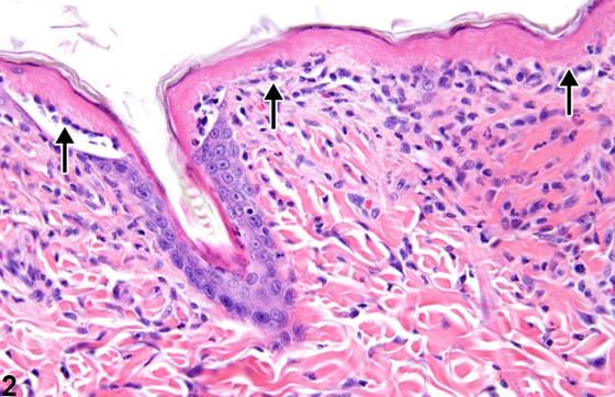
在皮肤毒性研究中,对毒性物质的初始反应通常涉及一定程度的上皮细胞损伤或破坏,如变性或坏死。在NTP研究中,作为原发性病变的上皮坏死(Figure 1、Figure 2)较溃疡形成报道少见。在严重病例中,上皮坏死可延伸至毛囊(Figure 3、Figure 4)。上皮坏死和毛囊坏死的特征为角质形成细胞皱缩或肿胀,胞质呈强嗜酸性至玻璃样变,细胞核浓缩,伴有不同程度的核固缩、核溶解和核碎裂。皮肤坏死(Figure 5、Figure 6)的特征为透壁性坏死,累及表皮、真皮,并可能累及皮下组织。作为原发性病变诊断者少见。坏死最常继发于表皮损伤,并常延伸至真皮和皮下组织。延伸至皮下组织的坏死不应与脂肪坏死混淆,后者局限于皮下组织,作为背景性病变散发性发生于小鼠和大鼠腹部皮下组织。营养不良性矿化常作为皮肤坏死的组成部分出现。转移性矿化与坏死或炎症无关,而是由钙磷失衡所致。
诊断与分级建议(NTP 共识)
观察到上皮坏死时,应作出诊断并赋予严重程度分级。在较严重病例中,毛囊上皮也可发生坏死;这应反映在赋予上皮坏死的严重程度分级中,并在病理学描述中予以描述。若毛囊坏死发生而无表皮受累,应使用术语“皮肤,毛囊 – 坏死”。相关病变,如炎症和溃疡形成,应单独诊断。皮肤坏死出现时,应记录其发生率并赋予严重程度分级。营养不良性矿化无需单独诊断,但应在病理学描述中予以描述。转移性矿化出现时应作出诊断,赋予严重程度分级,并在病理学描述中予以描述。脂肪坏死(即局限于皮下组织)应作为散发性背景性发现作出诊断、分级,并在描述中描述为“皮肤,皮下组织 – 坏死”。
图谱
英文原文
展开 NTP 官方英文描述与建议
Comment:
The initial response to a toxic agent in dermal toxicity studies usually involves some degree of epithelial cell damage or destruction, such as degeneration or necrosis. Epithelial necrosis ( Figure 1 and Figure 2 ) as a primary lesion is less frequently reported than ulceration in NTP studies. In severe cases, necrosis of the epithelium may extend into the hair follicles ( Figure 3 and Figure 4 ). Epithelial necrosis and follicular necrosis are characterized by shrunken or swollen keratinocytes with hypereosinophilic to hyalinized cytoplasm and condensed nuclei with varying degrees of pyknosis, karyolysis, and karyorrhexis. Skin necrosis ( Figure 5 and Figure 6 ) is characterized by transmural necrosis affecting the epidermis, dermis, and possibly the subcutis. It is infrequently diagnosed as a primary lesion. Necrosis most commonly occurs secondary to damage of the epidermis and frequently extends into the dermis and subcutis. Necrosis extending into the subcutis is not to be confused with fat necrosis, which is limited to the subcutis and occurs sporadically as a background lesion in the subcutaneous tissue of the abdomen in mice and rats. Dystrophic mineralization is commonly seen as a component of skin necrosis. Metastatic mineralization is not associated with necrosis or inflammation but due to calcium:phosphorus imbalance.
Recommendation:
When observed, epithelial necrosis should be diagnosed and assigned a severity grade. In more severe cases, the follicular epithelium may also be necrotic; this should be reflected in the severity grade assigned to epithelial necrosis and described in the pathology narrative. If follicular necrosis occurs without epidermal involvement, the term “Skin, Hair follicle – Necrosis” should be used. Associated lesions, such as inflammation and ulceration, should be diagnosed separately. Whenever present, the incidence of skin necrosis should be documented and assigned a severity grade. Dystrophic mineralization need not be diagnosed separately but should be described in the pathology narrative. Metastatic mineralization should be diagnosed whenever present, assigned a severity grade, and described in the pathology narrative. Fat necrosis (i.e., limited to the subcutis) should be diagnosed, graded, and described as a sporadic background finding in the narrative as “Skin, Subcutis – Necrosis.”
来源与引用
原始条目:https://ntp.niehs.nih.gov/atlas/nnl/integumentary-system/skin/Necrosis
图谱总目录:NTP Nonneoplastic Lesion Atlas。 内容属美国联邦政府作品(公有领域),可引用;引用时请标注来源。
建议引用格式:NTP Nonneoplastic Lesion Atlas. Necrosis (skin). LabSmart 中文版:
https://www.labsmart.cn/ntp/skin-necrosis.html
中文图谱总页(可展开对照全部 595 条):https://www.labsmart.cn/ntp-atlas.html#skin-necrosis