炎症Inflammation · 皮肤 · 皮肤系统 · NTP ntp-0372
诊断要点:无论何时出现,均应记录炎症的具体类型并赋予严重程度分级。当炎症作为与肿瘤、脂肪坏死或上皮坏死、或上皮囊肿或其他类型囊肿相关的继发性发现而存在时,炎症无需单独诊断,但应在病理学描述中予以描述。
病变描述
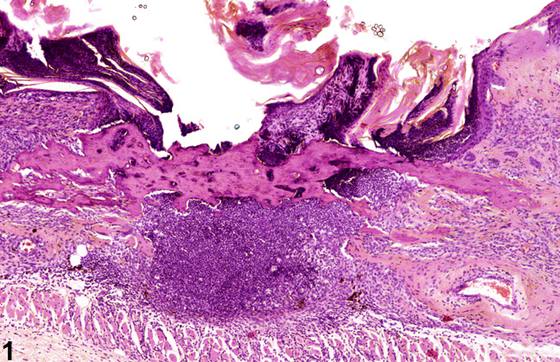
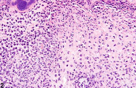
在皮肤给药毒性/致癌性研究中,炎症是对化学物质最常见的反应之一。炎症可能是非特异性的,也可能与自我损伤(抓挠)造成的创伤、接触设备边缘、节肢动物寄生或打斗造成的伤口有关。继发性炎症还可与皮肤肿瘤相关。皮肤急性炎症(Figure 1、Figure 2、Figure 3)的特征是中性粒细胞浸润,可伴有嗜酸性粒细胞和巨噬细胞,以及偶见的肥大细胞、淋巴细胞和浆细胞。化脓性炎症的特征是离散的退化中性粒细胞和细胞碎片聚集灶(Figure 4、Figure 5)。这些聚集灶周围还可见慢性化的证据,如纤维化和淋巴浆细胞浸润。慢性炎症(Figure 6、Figure 7)的特征是存在单核细胞(淋巴细胞、巨噬细胞和浆细胞),并可伴有纤维化或真皮细胞内黑色素蓄积。慢性活动性炎症(Figure 8、Figure 9)的特征是慢性炎症成分(淋巴细胞、巨噬细胞和纤维化)与叠加的急性炎症(中性粒细胞性和/或嗜酸性粒细胞性)并存。肉芽肿性炎症(Figure 10、Figure 11、Figure 12)的特征是巨噬细胞和多核巨细胞聚集,伴有数量不等的淋巴细胞、浆细胞或中性粒细胞。肉芽肿的一种典型形式是皮肤天然产物(如角蛋白、毛干、附件分泌物)通常因创伤而被引入结缔组织。这些皮肤产物作为异物,引发“异物性肉芽肿”,其特征性表现为多核巨细胞和/或上皮样巨噬细胞。所有形式的炎症均可伴有相关病变,包括水肿(尽管水肿更常与急性炎症相关)、上皮增生、角化过度、新生血管形成或出血,伴或不伴含含铁血黄素的巨噬细胞。
诊断与分级建议(NTP 共识)
无论何时出现,均应记录炎症的具体类型并赋予严重程度分级。当炎症作为与肿瘤、脂肪坏死或上皮坏死、或上皮囊肿或其他类型囊肿相关的继发性发现而存在时,炎症无需单独诊断,但应在病理学描述中予以描述。当存在水肿、出血或骨化生等继发性发现时,无需诊断,但应在描述中予以描述。如果炎症伴有上皮增生、角化过度或过度纤维化,此类发现应独立于慢性炎症进行诊断和分级。当肉芽肿性炎症伴有异物时,两种诊断应分别记录,但异物诊断不应分级。
图谱
英文原文
展开 NTP 官方英文描述与建议
Comment:
In dermal application toxicity/carcinogenicity studies, inflammation is one of the most common responses to chemicals. Inflammation may be nonspecific or related to trauma from self-mutilation (scratching), contact with edges of equipment, arthropod parasitism, or wounds from fighting. Secondary inflammation can also be associated with cutaneous neoplasms. Acute inflammation ( Figure 1 , Figure 2 , and Figure 3 ) in the skin is characterized by infiltration of neutrophils, which may be accompanied by eosinophils and macrophages as well as occasional mast cells, lymphocytes, and plasma cells. Suppurative inflammation is characterized by discrete pockets of degenerate neutrophils and cellular debris ( Figure 4 and Figure 5 ). Evidence of chronicity, such as fibrosis and lymphoplasmacytic infiltrates, may also surround these pockets. Chronic inflammation ( Figure 6 and Figure 7 ) is characterized by the presence of mononuclear cells (lymphocytes, macrophages, and plasma cells) and may be accompanied by fibrosis or the accumulation of melanin within cells in the dermis. Chronic active inflammation ( Figure 8 and Figure 9 ) is characterized by the coexistence of elements of chronic inflammation (lymphocytes, macrophages, and fibrosis) and superimposed acute inflammation (neutrophilic and/or eosinophilic). Granulomatous inflammation ( Figure 10 , Figure 11 , and Figure 12 ) is characterized by an accumulation of macrophages and multinucleated giant cells, with variable numbers of lymphocytes, plasma cells, or neutrophils. A typical form of granuloma is one in which natural products of the skin (e.g., keratin, hair shaft, adnexal secretions) are introduced into the connective tissues, usually by trauma. These skin products act as foreign bodies, inciting a “foreign body granuloma,” a characteristic feature of which are multinucleated giant cells and/or epithelioid macrophages. All forms of inflammation may be accompanied by associated lesions, including edema (though this is more commonly associated with acute inflammation), epithelial hyperplasia, hyperkeratosis, neovascularization, or hemorrhage with or without hemosiderin-containing macrophages.
Recommendation:
Whenever present, the specific type of inflammation should be recorded and assigned a severity grade. When present as a secondary finding associated with neoplasia, fat necrosis or epithelial necrosis, or epithelial or other types of cysts, inflammation need not be diagnosed separately but should be described in the pathology narrative. When secondary findings such as edema, hemorrhage, or osseous metaplasia are present, they need not be diagnosed but should be described in the narrative. If the inflammation is accompanied by epithelial hyperplasia, hyperkeratosis, or excessive fibrosis, such findings should be diagnosed and graded independently of chronic inflammation. When granulomatous inflammation is accompanied by foreign material, both diagnoses should be documented separately, but the diagnosis of foreign material should not be graded.
来源与引用
原始条目:https://ntp.niehs.nih.gov/atlas/nnl/integumentary-system/skin/Inflammation
图谱总目录:NTP Nonneoplastic Lesion Atlas。 内容属美国联邦政府作品(公有领域),可引用;引用时请标注来源。
建议引用格式:NTP Nonneoplastic Lesion Atlas. Inflammation (skin). LabSmart 中文版:
https://www.labsmart.cn/ntp/skin-inflammation.html
中文图谱总页(可展开对照全部 595 条):https://www.labsmart.cn/ntp-atlas.html#skin-inflammation