色素Pigment · 皮肤 · 皮肤系统 · NTP ntp-0376
诊断要点:在有指征时(根据病理学家的判断),色素应当被记录并赋予严重程度分级。如果色素是炎症的次要成分,则不必单独诊断,但应在叙述中描述。
病变描述
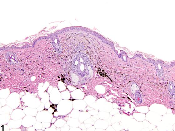
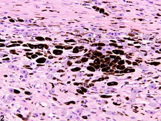
皮肤色素的诊断以真皮或皮下组织中载色素细胞(黑素细胞或巨噬细胞)的蓄积为特征(Figure 1、Figure 2)。色素几乎仅见于皮肤给药研究,是局部涂敷受试物后反应的一部分。因此,色素通常伴随其他病变,如炎症(通常为慢性或慢性活动性)或纤维化。
诊断与分级建议(NTP 共识)
在有指征时(根据病理学家的判断),色素应当被记录并赋予严重程度分级。如果色素是炎症的次要成分,则不必单独诊断,但应在叙述中描述。
图谱
英文原文
展开 NTP 官方英文描述与建议
Comment:
The diagnosis of pigment in the skin is characterized by the accumulation of pigment-laden cells (melanocytes or macrophages) in the dermis or subcutis ( Figure 1 and Figure 2 ). Pigment occurs almost exclusively in dermal studies as part of the response to topical application of a test article. Hence, pigment is typically accompanied by other lesions, such as inflammation (usually chronic or chronic active) or fibrosis.
Recommendation:
When warranted (based on the judgment of the pathologist), pigment should be documented and assigned a severity grade. If it is a minor component of inflammation, it need not be diagnosed separately but should be described in the narrative.
来源与引用
原始条目:https://ntp.niehs.nih.gov/atlas/nnl/integumentary-system/skin/Pigment
图谱总目录:NTP Nonneoplastic Lesion Atlas。 内容属美国联邦政府作品(公有领域),可引用;引用时请标注来源。
建议引用格式:NTP Nonneoplastic Lesion Atlas. Pigment (skin). LabSmart 中文版:
https://www.labsmart.cn/ntp/skin-pigment.html
中文图谱总页(可展开对照全部 595 条):https://www.labsmart.cn/ntp-atlas.html#skin-pigment