滤泡扩张Follicle Dilation · 甲状腺 · 内分泌系统 · NTP ntp-0064
诊断要点:推荐对扩张滤泡进行记录和分级。诊断应指明扩张是否为双侧性,严重程度分级应基于受累更严重的甲状腺。
病变描述
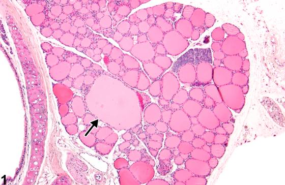
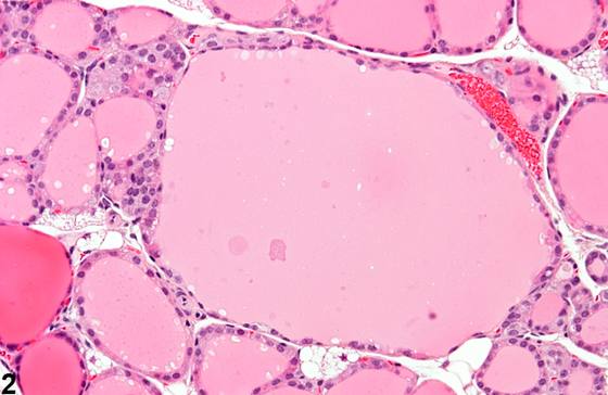
一个或多个扩张的滤泡是小鼠和大鼠甲状腺中常见的、与年龄相关的改变。受累滤泡的胶质可呈淡染(Figure 1、Figure 2)或缺失,管腔内可见脱落细胞。扩张的滤泡常见于甲状腺外周(Figure 3、Figure 4)。扩张的滤泡衬以低立方至扁平上皮。
诊断与分级建议(NTP 共识)
推荐对扩张滤泡进行记录和分级。诊断应指明扩张是否为双侧性,严重程度分级应基于受累更严重的甲状腺。扩张滤泡管腔内出现细胞无需单独诊断,除非其严重程度足以需要单独诊断。
图谱
英文原文
展开 NTP 官方英文描述与建议
Comment:
One or more dilated follicles occur as a common and age-associated change in mouse and rat thyroids. Affected follicles may have pale ( Figure 1 and Figure 2 ) or absent colloid, and exfoliated cells may be present in the lumen. The dilated follicles are frequently seen at the periphery of the thyroid gland ( Figure 3 and Figure 4 ). Dilated follicles are lined by low cuboidal to flattened epithelium.
Recommendation:
Documentation and grading of dilated follicles are recommended. The diagnosis should indicate whether the dilation is bilateral, with a severity grade based on the more severely affected thyroid gland. The presence of cells within the lumens of the dilated follicles need not be diagnosed separately unless warranted by severity.
来源与引用
原始条目:https://ntp.niehs.nih.gov/atlas/nnl/endocrine-system/thyroid-gland/Follicle-Dilation
图谱总目录:NTP Nonneoplastic Lesion Atlas。 内容属美国联邦政府作品(公有领域),可引用;引用时请标注来源。
建议引用格式:NTP Nonneoplastic Lesion Atlas. Follicle Dilation (thyroid-gland). LabSmart 中文版:
https://www.labsmart.cn/ntp/thyroid-gland-follicle-dilation.html
中文图谱总页(可展开对照全部 595 条):https://www.labsmart.cn/ntp-atlas.html#thyroid-gland-follicle-dilation