滤泡上皮增生Follicle Epithelium Hyperplasia · 甲状腺 · 内分泌系统 · NTP ntp-0062
诊断要点:尽管滤泡增生可自发发生,但一旦存在即应作出诊断并给予严重程度分级。严重程度可依据受累腺体的百分比及增生的形态学复杂程度判定。
病变描述
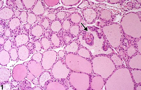
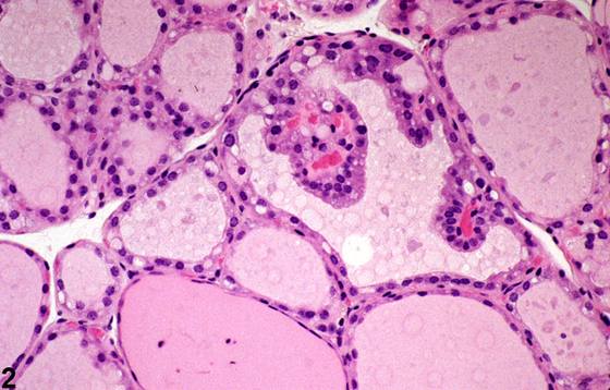
滤泡上皮增生(hyperplasia of follicular epithelium)可为局灶性(focal)、弥漫性(diffuse),有时呈囊性。局灶性或多灶性(multifocal)滤泡增生可存在于单个或少数几个滤泡中。局灶性增生无包膜,不压迫周围实质,借此与良性肿瘤相鉴别。受累滤泡可较小、含极少量胶质,或较大而不规则,增生的上皮呈乳头状突起伸入滤泡腔(Figure 1、Figure 2)。甲状腺滤泡肥大(hypertrophy)与增生常同时发生(Figure 3)。亦可出现以滤泡上皮细胞核拥挤、向滤泡腔内突起极轻微为特征的增生(Figure 4)。
诊断与分级建议(NTP 共识)
尽管滤泡增生可自发发生,但一旦存在即应作出诊断并给予严重程度分级。严重程度可依据受累腺体的百分比及增生的形态学复杂程度判定。增生的性质与范围以及为单侧抑或双侧,均应在病理描述中予以说明。建议区分局灶性(focal)、弥漫性(diffuse)与囊性甲状腺滤泡增生,并应将其作为诊断的限定词纳入诊断。伴增生的滤泡中任何相关肥大的性质不应单独诊断,除非其严重程度足以支持单独诊断。若未作诊断,则应在病理描述中说明肥大情况。
图谱
英文原文
展开 NTP 官方英文描述与建议
Comment:
Hyperplasia of follicular epithelium may be focal, diffuse, or sometimes cystic. Focal or multifocal follicular hyperplasia can be present in a single or in a few follicles. Focal hyperplasia is not encapsulated and does not compress surrounding parenchyma, which differentiates it from benign tumors. Affected follicles may be small with minimal colloid or large and irregular with papillary projections of hyperplastic epithelium extending into the follicular lumen ( Figure 1 and Figure 2 ). Thyroid follicular hypertrophy and hyperplasia often occur together ( Figure 3 ). Hyperplasia characterized by crowding of follicular epithelium nuclei and minimal protrusion into the follicular lumen may occur ( Figure 4 ).
Recommendation:
Although follicular hyperplasia can occur spontaneously, it should be diagnosed and given a severity grade whenever present. Severity may be based on the percentage of the gland involved and the morphologic complexity of the hyperplasia. The nature and extent of the hyperplasia and whether it is unilateral or bilateral should be described in the pathology narrative. Distinction between focal, diffuse, and cystic thyroid follicular hyperplasia is recommended and should be included as a qualifier of the diagnosis. The nature of any associated hypertrophy in follicles with hyperplasia should not be diagnosed separately, unless warranted by severity. If not diagnosed, hypertrophy should be described in the pathology narrative.
来源与引用
原始条目:https://ntp.niehs.nih.gov/atlas/nnl/endocrine-system/thyroid-gland/FollicleEpithelium-Hyperplasia
图谱总目录:NTP Nonneoplastic Lesion Atlas。 内容属美国联邦政府作品(公有领域),可引用;引用时请标注来源。
建议引用格式:NTP Nonneoplastic Lesion Atlas. Follicle Epithelium Hyperplasia (thyroid-gland). LabSmart 中文版:
https://www.labsmart.cn/ntp/thyroid-gland-follicle-epithelium-hyperplasia.html
中文图谱总页(可展开对照全部 595 条):https://www.labsmart.cn/ntp-atlas.html#thyroid-gland-follicle-epithelium-hyperplasia