结膜水肿Conjunctiva Edema · 眼 · 特殊感觉系统 · NTP ntp-0336
诊断要点:结膜水肿应当被诊断并赋予严重程度分级。如果位于球结膜,水肿可在器官/亚部位“眼,结膜”下诊断。
病变描述
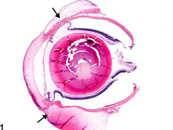
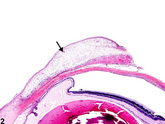
球结膜水肿(Figure 1、Figure 2、Figure 3)的特征是清亮至淡嗜酸性液体积聚所致的弥漫性肿胀。临床上,结膜水肿称为 chemosis。它是一种非特异性病变,继发于局部刺激物、变应原、创伤和感染因子等原因。水肿区域可能存在炎性细胞。
诊断与分级建议(NTP 共识)
结膜水肿应当被诊断并赋予严重程度分级。如果位于球结膜,水肿可在器官/亚部位“眼,结膜”下诊断。如果位于睑结膜,则可在“皮肤,眼睑”的器官/亚部位下诊断。如果继发于另一过程(例如炎症),结膜水肿不应单独诊断(除非严重程度确有必要),而应在叙述中描述。
图谱
英文原文
展开 NTP 官方英文描述与建议
Comment:
Edema of the bulbar conjunctiva ( Figure 1 , Figure 2 , and Figure 3 ) is characterized by diffuse swelling due to accumulation of clear to pale eosinophilic fluid. Clinically, conjunctival edema is referred to as chemosis. It is a nonspecific finding secondary to such causes as topical irritants, allergens, trauma, and infectious agents. Inflammatory cells may be present in the edematous areas.
Recommendation:
Conjunctival edema should be diagnosed and assigned a severity grade. If located in the bulbar conjunctiva, edema can be diagnosed under the organ/subtopography "eye, conjunctiva." If located in the palpebral conjunctiva, it can be diagnosed under the organ/subtopography of “skin, eyelid.” If secondary to another process (e.g., inflammation), conjunctival edema should not be diagnosed separately (unless warranted by severity) but should be described in the narrative.
来源与引用
原始条目:https://ntp.niehs.nih.gov/atlas/nnl/special-senses-system/eye/Conjunctiva-Edema
图谱总目录:NTP Nonneoplastic Lesion Atlas。 内容属美国联邦政府作品(公有领域),可引用;引用时请标注来源。
建议引用格式:NTP Nonneoplastic Lesion Atlas. Conjunctiva Edema (eye). LabSmart 中文版:
https://www.labsmart.cn/ntp/eye-conjunctiva-edema.html
中文图谱总页(可展开对照全部 595 条):https://www.labsmart.cn/ntp-atlas.html#eye-conjunctiva-edema