角膜细胞质空泡化Cornea Vacuolation Cytoplasmic · 眼 · 特殊感觉系统 · NTP ntp-0353
诊断要点:如果作为原发性毒性或遗传性效应发生,角膜细胞质空泡化应予以诊断并赋予严重程度分级。
病变描述
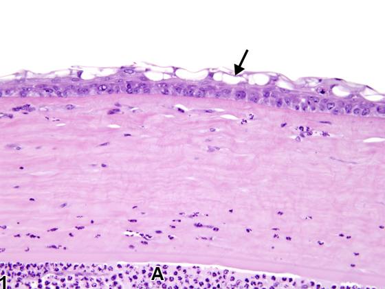
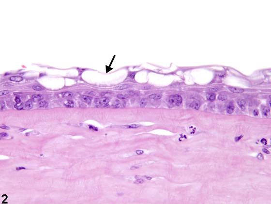
角膜上皮空泡(Figure 1、Figure 2、Figure 3)的特征为离散、大小不一、通常清亮、单个至多个、圆形至卵形的细胞质空腔。这些空泡可发生于角膜上皮细胞、内皮细胞或基质细胞(角膜细胞)。上皮空泡可发生于特定的细胞层(例如 basal、suprabasal)。空泡可增大并融合,形成上皮内囊泡或基质腔隙。角膜细胞中的空泡可由多种原因引起,包括全身性或局部应用的毒物;可作为遗传性贮积病(如黏多糖贮积症)的特征出现;或可继发于角膜干燥或炎症。真性空泡必须与角膜细胞中常出现的伪影性清亮区(常为核周)相鉴别。空泡化可伴有下方基质(Figure 1、Figure 2、Figure 3)或前房(Figure 1)中的炎症细胞,以及角膜上皮增生和角化过度(Figure 3)。
诊断与分级建议(NTP 共识)
如果作为原发性毒性或遗传性效应发生,角膜细胞质空泡化应予以诊断并赋予严重程度分级。诊断中应包含适当的部位修饰词(例如 epithelium、endothelium、stroma),以指示受累细胞的位置。当作为另一病理过程(例如炎症)的特征或后遗症发生时,角膜细胞质空泡化不应单独诊断,除非严重程度足以支持,但应在病理学叙述中描述。相关链接:Eye - Cornea - Edema
图谱
英文原文
展开 NTP 官方英文描述与建议
Comment:
Corneal epithelial vacuoles ( Figure 1 , Figure 2 , and Figure 3 ) are characterized as discrete, variably sized, generally clear, single to multiple, round to oval cytoplasmic spaces. These can occur in the corneal epithelial, endothelial, or stromal cells (keratocytes). Epithelial vacuoles can occur in specific cell layers (e.g., basal, suprabasal). Vacuoles may enlarge and coalesce to form intraepithelial vesicles or stromal lacunae. Vacuoles in corneal cells can result from various causes, including systemic or topically applied toxins; can appear as features of inherited storage diseases, such as mucopolysaccharidoses; or can be secondary to corneal desiccation or inflammation. True vacuoles must be distinguished from the artifactual clear zones (often perinuclear) that often occur in corneal cells. The vacuolation may be accompanied by inflammatory cells in the underlying stroma ( Figure 1 , Figure 2 , and Figure 3 ) or the anterior chamber ( Figure 1 ), and corneal epithelial hyperplasia and hyperkeratosis ( Figure 3 ).
Recommendation:
If occurring as a primary toxic or heritable effect, corneal cytoplasmic vacuolation should be diagnosed and assigned a severity grade. An appropriate site modifier (e.g., epithelium, endothelium, stroma) should be included in the diagnosis to indicate the location of the affected cells. When occurring as a feature or sequela to another pathologic process (e.g., inflammation), corneal cytoplasmic vacuolation should not be diagnosed separately unless warranted by severity, but should be described in the pathology narrative. Related Links: Eye - Cornea - Edema
来源与引用
原始条目:https://ntp.niehs.nih.gov/atlas/nnl/special-senses-system/eye/Cornea-VacuolationCytoplasmic
图谱总目录:NTP Nonneoplastic Lesion Atlas。 内容属美国联邦政府作品(公有领域),可引用;引用时请标注来源。
建议引用格式:NTP Nonneoplastic Lesion Atlas. Cornea Vacuolation Cytoplasmic (eye). LabSmart 中文版:
https://www.labsmart.cn/ntp/eye-cornea-vacuolation-cytoplasmic.html
中文图谱总页(可展开对照全部 595 条):https://www.labsmart.cn/ntp-atlas.html#eye-cornea-vacuolation-cytoplasmic