视神经变性Optic Nerve Degeneration · 眼 · 特殊感觉系统 · NTP ntp-0338
诊断要点:视神经变性应当诊断并赋予严重程度分级。视神经变性的存在应当促使仔细检查视网膜(尤其是神经节细胞层和神经纤维层)以发现并发病理改变。
病变描述
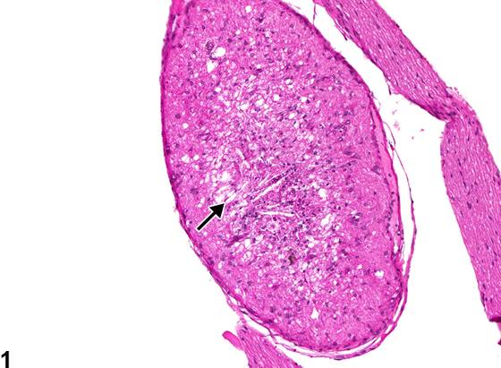
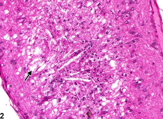
视神经变性(optic nerve degeneration)可由多种原因引起,包括眼内压升高、血管供应受损以及物理性创伤(例如来自眶后采血操作)。由于视神经由视网膜神经节细胞的轴突构成,视网膜的原发性病变(尤其是影响神经节细胞层和神经纤维层的病变)常导致并发的视神经病理改变,包括变性。由于视神经是脑的颅外白质束,视神经变性也可以是继发于脑部病变的“下行性”改变。尽管程度可随啮齿动物品系而异,视神经轴突变性和丢失也可以是大鼠和小鼠的年龄相关偶然性改变。病因不明的视神经变性也有报道为成年大鼠的自发性改变。视神经变性的特征可为散在的空泡(如 Figure 1、Figure 2 所示),其中常含有肿胀、淡染的嗜酸性粒细胞(eosinophil)碎片化轴突和/或髓鞘碎片。在更慢性的病例中,可出现轴突数量减少、残留轴突直径减小以及软膜隔增厚。轴突丢失可导致神经整体直径减小。可同时发生程度不等的胶质细胞增生和/或巨噬细胞浸润。
诊断与分级建议(NTP 共识)
视神经变性应当诊断并赋予严重程度分级。视神经变性的存在应当促使仔细检查视网膜(尤其是神经节细胞层和神经纤维层)以发现并发病理改变。相关病变(例如炎症)应当单独诊断。相关链接:Eye - Optic Nerve - Gliosis
图谱
英文原文
展开 NTP 官方英文描述与建议
Comment:
Optic nerve degeneration can result from various causes, including increased intraocular pressure, compromised vascular supply, and physical trauma (e.g., from retro-orbital bleeding procedures). Since the optic nerve is composed of the axons of retinal ganglion cells, primary lesions in the retina (especially those affecting the ganglion cell and nerve fiber layers) frequently result in concurrent optic nerve pathology, including degeneration. Since the optic nerve is an extracranial white matter tract of the brain, optic nerve degeneration can also be a "descending" change secondary to lesions in the brain. Although the extent can vary with rodent strain, optic nerve axonal degeneration and loss can also be an age-related incidental change in rats and mice. Optic nerve degeneration of uncertain etiology has also been reported as a spontaneous change in adult rats. Optic nerve degeneration can be characterized by scattered clear vacuoles (as in Figure 1 and Figure 2 ), which often contain swollen, pale eosinophilic fragmented axons and/or myelin debris. In more chronic cases, there can be decreased numbers of axons, decreased diameter of the remaining axons, and pial septa thickening. The loss of axons may lead to decreased overall diameter of the nerve. Variable glial cell proliferation and/or macrophage infiltration may occur concurrently.
Recommendation:
Optic nerve degeneration should be diagnosed and assigned a severity grade. The presence of optic nerve degeneration should prompt careful examination of the retina (especially the ganglion cell and nerve fiber layers) for concurrent pathology. Associated lesions (e.g., inflammation) should be diagnosed separately. Related Links: Eye - Optic Nerve - Gliosis
来源与引用
原始条目:https://ntp.niehs.nih.gov/atlas/nnl/special-senses-system/eye/OpticNerve-Degeneration
图谱总目录:NTP Nonneoplastic Lesion Atlas。 内容属美国联邦政府作品(公有领域),可引用;引用时请标注来源。
建议引用格式:NTP Nonneoplastic Lesion Atlas. Optic Nerve Degeneration (eye). LabSmart 中文版:
https://www.labsmart.cn/ntp/eye-optic-nerve-degeneration.html
中文图谱总页(可展开对照全部 595 条):https://www.labsmart.cn/ntp-atlas.html#eye-optic-nerve-degeneration