眼球萎缩Phthisis Bulbi · 眼 · 特殊感觉系统 · NTP ntp-0335
诊断要点:眼球痨应诊断为存在,但不进行严重程度分级。眼部各亚部位的变化不应单独诊断,因为它们已包含在集合性诊断术语“眼球痨”中。
病变描述
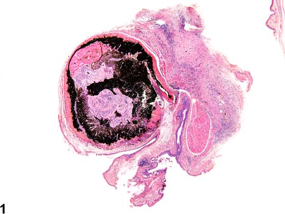
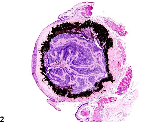
眼球痨(phthisis bulbi)(Figure 1、Figure 2、Figure 3、Figure 4)是用于描述“终末期”眼球形态的诊断术语,该形态由多种原因所致的严重损伤引起,例如创伤、炎症、电离辐射或环境光过度暴露,或眶内占位性肿块(如哈德氏腺肿瘤)的物理压迫(Figure 4)。眼球痨的特征是眼球显著皱缩,伴有眼结构的明显移位和结构紊乱。
诊断与分级建议(NTP 共识)
眼球痨应诊断为存在,但不进行严重程度分级。眼部各亚部位的变化不应单独诊断,因为它们已包含在集合性诊断术语“眼球痨”中。如果存在诱发原因(例如异物),应单独诊断,并应在叙述中描述这两种病变之间的关联。
图谱
英文原文
展开 NTP 官方英文描述与建议
Comment:
Phthisis bulbi ( Figure 1 , Figure 2 , Figure 3 , and Figure 4 ) is the diagnostic term used for the "end-stage" eye morphology resulting from severe injury from various causes, such as trauma, inflammation, excess exposure to ionizing radiation or ambient light, or physical compression by space-occupying orbital masses, such as neoplasms of the Harderian gland ( Figure 4 ). Phthisis bulbi is characterized by prominent shrinkage of the globe with marked displacement and disorganization of ocular structures.
Recommendation:
Phthisis bulbi should be diagnosed as present without assignment of a severity grade. Changes in the various ocular subtopographies should not be diagnosed separately as they are included in the collective diagnostic term "phthisis bulbi." If an inciting cause (e.g., a foreign body) is present, it should be diagnosed separately, and the association between the two lesions should be described in the narrative.
来源与引用
原始条目:https://ntp.niehs.nih.gov/atlas/nnl/special-senses-system/eye/PhthisisBulbi
图谱总目录:NTP Nonneoplastic Lesion Atlas。 内容属美国联邦政府作品(公有领域),可引用;引用时请标注来源。
建议引用格式:NTP Nonneoplastic Lesion Atlas. Phthisis Bulbi (eye). LabSmart 中文版:
https://www.labsmart.cn/ntp/eye-phthisis-bulbi.html
中文图谱总页(可展开对照全部 595 条):https://www.labsmart.cn/ntp-atlas.html#eye-phthisis-bulbi