视网膜变性Retina Degeneration · 眼 · 特殊感觉系统 · NTP ntp-0342
诊断要点:视网膜变性只要存在即应诊断并分级。应在病理学描述中描述病变在视网膜内的位置(周边或中央)和范围,以及受累的视网膜各层。
病变描述
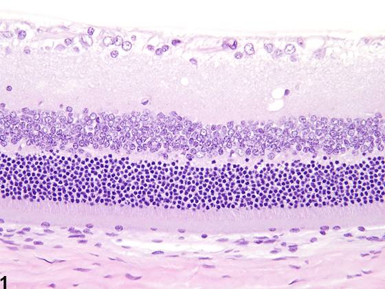
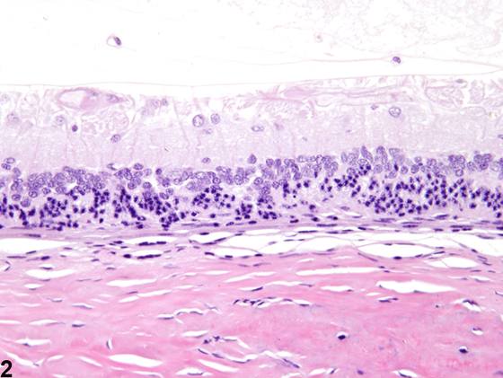
大鼠和小鼠的视网膜变性可作为衰老性改变发生,也可继发于多种损伤,如物理性创伤、视网膜脱离、炎症、感染性病原体(例如病毒)、血管紊乱(例如梗死)、眼内压升高和营养缺乏。在许多实验室小鼠和大鼠品系中也已记录到多种遗传性视网膜变性。当大鼠和小鼠(尤其是白化品系)暴露于过强的环境光照时,可发生所谓的光诱导性视网膜变性。视网膜变性也可为全身或局部给予化学物的直接毒性效应。在实验室大鼠和小鼠中也有病因不明的自发性视网膜变性报道。在许多类型的视网膜变性中,最常受累的视网膜神经元是视杆和视锥光感受器及/或神经节细胞。根据病因不同,退行性改变可局限于(或更广泛地分布于)特定的视网膜拓扑区域(即上方与下方、中央与周边)。轻度视网膜变性(Figure 2,与Figure 1比较)通常由视杆和视锥光感受器突丢失、外核层光感受器细胞凋亡、内、外核层细胞减少和结构紊乱,以及丛状层变窄或消失组成。更明显的视网膜变性(Figure 3,与Figure 1比较)通常以整体变薄、结构破坏和细胞成分丢失,以及视杆和视锥光感受器及丛状层完全消失为特征。变性视网膜还可表现出其他形态学特征,如视网膜胶质细胞(例如Müller细胞和星形胶质细胞)的弥漫性(diffuse)或nodular增生、固定皱褶和菊形团样结构、视网膜内腔隙或囊肿(“微囊样”形成和视网膜劈裂),以及相邻视网膜色素上皮的伴发性增生和/或视网膜内迁移。大鼠和小鼠还可表现出一种称为周边视网膜变性的偶然性衰老改变(与比较),其特征为由于细胞减少或视网膜各层缺失所致的周边视网膜显著(marked)变薄。尽管未附图说明,囊性腔隙(“微囊样”改变)也是这种周边年龄相关变性的常见特征。
诊断与分级建议(NTP 共识)
视网膜变性只要存在即应诊断并分级。应在病理学描述中描述病变在视网膜内的位置(周边或中央)和范围,以及受累的视网膜各层。病理学家关于该病变是周边性、年龄相关性、背景性病变(或其加重)还是原发性治疗相关病变的意见应纳入病理学描述。视网膜变性的各种形态学特征(特定层的细胞减少、视杆和视锥光感受器突丢失等)不应单独诊断,而应在描述中加以描述。视网膜变性的存在应促使仔细检查视神经以发现伴发性病变。
图谱
英文原文
展开 NTP 官方英文描述与建议
Comment:
Retinal degeneration in rats and mice can occur as an aging change or secondary to various insults, such as physical trauma, retinal detachment, inflammation, infectious agents (e.g., viruses), vascular derangements (e.g., infarction), increased intraocular pressure, and nutritional deficiencies. Numerous heritable retinal degenerations have also been documented in various strains of laboratory mice and rats. So-called light-induced retinal degeneration can occur when rats and mice (especially albino strains) are exposed to overly intense ambient light. Retinal degeneration can also be a direct toxic effect of systemically or topically administered chemical agents. Spontaneously occurring retinal degeneration of uncertain etiology has also been reported in laboratory rats and mice. In many types of retinal degeneration, the most commonly affected retinal neurons are the rod and cone photoreceptors and/or the ganglion cells. Depending on the cause, degenerative changes may be localized (or more extensive) in particular retinal topographic regions (i.e., superior vs. inferior, central vs. peripheral). Mild retinal degeneration ( Figure 2 , compare to Figure 1 ) typically consists of loss of the rod and cone photoreceptor processes, apoptosis of outer nuclear layer photoreceptor cells, hypocellularity and disorganization of the inner and outer nuclear layers, and narrowing or absence of the plexiform layers. More pronounced retinal degeneration ( Figure 3 , compare to Figure 1 ) is typically characterized by overall thinning, architectural disruption, and loss of cellular elements, as well as complete absence of the rod and cone photoreceptors and the plexiform layers. Degenerate retinas may also exhibit other morphologic features, such as diffuse or nodular proliferation of retinal glial cells (e.g., Müller cells and astrocytes), fixed fold and rosette-like formations, and intraretinal cavitations or cysts ("microcystoid" formation and retinoschisis), as well as concurrent proliferation and/or intraretinal migration of the adjacent retinal pigment epithelium. Rats and mice can also exhibit an incidental aging change known as peripheral retinal degeneration ( and , compare to ), which is characterized by marked thinning of the peripheral retina due to hypocellularity or absence of retinal layers. Although not illustrated, cystic cavitations ("microcystoid" change) are also common features of this peripheral age-related degeneration.
Recommendation:
Retinal degeneration should be diagnosed and graded whenever present. The location (peripheral or central) and extent of the lesion within the retina, as well as the retinal layers affected, should be described in the pathology narrative. The pathologist's opinion as to whether it is the peripheral, age related, background form of the lesion (or an exacerbation thereof) or a primary treatment-related lesion should be included in the pathology narrative. The various morphologic features of retinal degeneration (hypocellularity of specific layers, rod and cone photoreceptor process loss, etc.) should not be diagnosed separately but should be described in the narrative. The presence of retinal degeneration should prompt careful examination of the optic nerve for concurrent lesions.
来源与引用
原始条目:https://ntp.niehs.nih.gov/atlas/nnl/special-senses-system/eye/Retina-Degeneration
图谱总目录:NTP Nonneoplastic Lesion Atlas。 内容属美国联邦政府作品(公有领域),可引用;引用时请标注来源。
建议引用格式:NTP Nonneoplastic Lesion Atlas. Retina Degeneration (eye). LabSmart 中文版:
https://www.labsmart.cn/ntp/eye-retina-degeneration.html
中文图谱总页(可展开对照全部 595 条):https://www.labsmart.cn/ntp-atlas.html#eye-retina-degeneration