视网膜胶质增生Retina Gliosis · 眼 · 特殊感觉系统 · NTP ntp-0343
诊断要点:当作为原发性改变发生时,视网膜胶质增生应当被诊断并赋予严重程度分级。当视网膜胶质增生作为另一病理过程(如视网膜变性)的形态学特征或反应性继发结果发生时,不应单独诊断(除非因严重程度而有必要),但应在病理学描述中予以描述。
病变描述
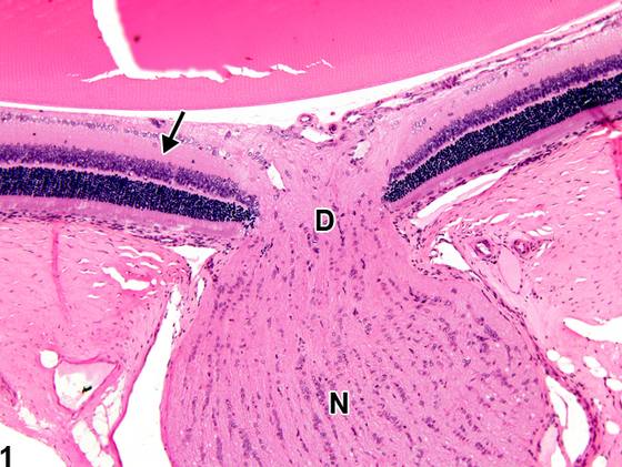
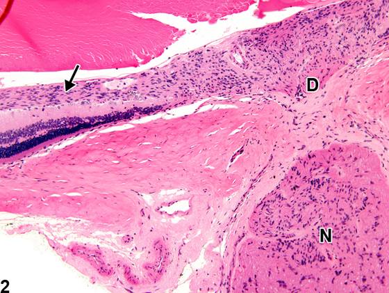
视网膜胶质增生(retinal gliosis,图2,与图1中的正常表现相比较),即星形胶质细胞(astrocytes)、Müller细胞和/或小胶质细胞(microglia)的增生,可发生于视网膜各层,呈局灶性(focal)至弥漫性(diffuse)分布。其特征为视网膜内胶质细胞数量增多。视网膜胶质增生可作为原发性改变(通常病因不明)发生,也可作为其他视网膜病变(如变性)的一种特征出现。
诊断与分级建议(NTP 共识)
当作为原发性改变发生时,视网膜胶质增生应当被诊断并赋予严重程度分级。当视网膜胶质增生作为另一病理过程(如视网膜变性)的形态学特征或反应性继发结果发生时,不应单独诊断(除非因严重程度而有必要),但应在病理学描述中予以描述。
图谱
英文原文
展开 NTP 官方英文描述与建议
Comment:
Retinal gliosis ( Figure 2 , compare to normal in Figure 1 ), the proliferation of astrocytes, Müller cells, and/or microglia, can occur in various retinal layers with focal to diffuse distribution. It is characterized by increased numbers of glial cells in the retina. Retinal gliosis can occur as a primary change (generally of uncertain etiology) or as a feature to other retinal lesions (e.g., degeneration).
Recommendation:
When occurring as a primary change, retinal gliosis should be diagnosed and assigned a severity grade. When retinal gliosis occurs as a morphologic feature or reactive sequela of another pathologic process (e.g., retinal degeneration), it should not be diagnosed separately (unless warranted by severity), but should be described in the pathology narrative.
来源与引用
原始条目:https://ntp.niehs.nih.gov/atlas/nnl/special-senses-system/eye/Retina-Gliosis
图谱总目录:NTP Nonneoplastic Lesion Atlas。 内容属美国联邦政府作品(公有领域),可引用;引用时请标注来源。
建议引用格式:NTP Nonneoplastic Lesion Atlas. Retina Gliosis (eye). LabSmart 中文版:
https://www.labsmart.cn/ntp/eye-retina-gliosis.html
中文图谱总页(可展开对照全部 595 条):https://www.labsmart.cn/ntp-atlas.html#eye-retina-gliosis