淋巴细胞凋亡Apoptosis Lymphocyte · 脾脏 · 免疫系统 · NTP ntp-0035
诊断要点:当脾脏淋巴细胞凋亡超过同期对照所见时,应当诊断并赋予严重程度分级。淋巴细胞凋亡必须与脾脏坏死相鉴别。
病变描述
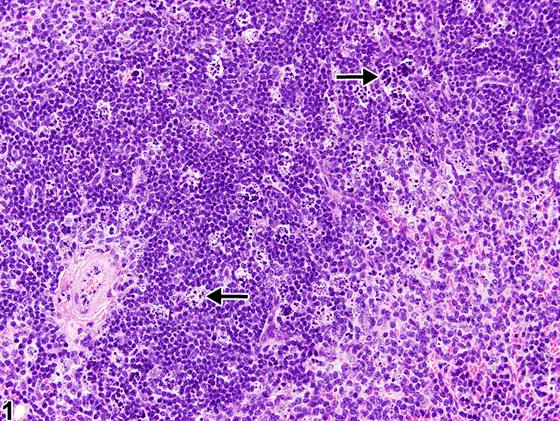
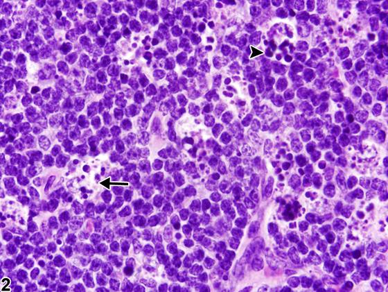
淋巴细胞凋亡(lymphocyte apoptosis)通常发生于啮齿动物脾脏富含B细胞的滤泡生发中心,但在实验性暴露于辐射、病毒、内毒素或化学物时,B细胞和/或T细胞(动脉周围淋巴鞘)区域内的淋巴细胞凋亡也可能增多。凋亡的特征是单个淋巴细胞皱缩、核染色质浓缩,以及凋亡细胞碎裂成膜包裹的凋亡小体(apoptotic bodies)(Figure 2,箭头),随后被巨噬细胞(可染体巨噬细胞,tingible body macrophages)吞噬(Figure 1、Figure 2,箭头)。显著的淋巴细胞凋亡可导致一个或多个白髓区域萎缩。历史上,淋巴细胞凋亡曾被称为“淋巴细胞坏死(lymphocyte necrosis)”,但该病变的正确术语是“淋巴细胞凋亡”。当主要细胞形态学符合经典坏死形式时,应使用坏死的诊断(见脾脏-坏死,Spleen - Necrosis)。坏死的特征是细胞肿胀、核浓缩、核碎裂和核溶解、细胞溶解,以及大量嗜酸性胞质和核碎裂碎屑的积聚。坏死通常伴有炎症。
诊断与分级建议(NTP 共识)
当脾脏淋巴细胞凋亡超过同期对照所见时,应当诊断并赋予严重程度分级。淋巴细胞凋亡必须与脾脏坏死相鉴别。相关链接:脾脏-坏死(Spleen - Necrosis)
图谱
英文原文
展开 NTP 官方英文描述与建议
Comment:
Lymphocyte apoptosis normally occurs in the B-cell-rich follicular germinal centers of the spleen of rodents but may also be increased in B-cell and/or T-cell (periarteriolar lymphatic sheaths) compartments with experimental exposures to radiation, viruses, endotoxin, or chemicals. Apoptosis is characterized by shrinkage of individual lymphocytes, condensation of nuclear chromatin, and fragmentation of apoptotic cells into membrane-bound bodies (apoptotic bodies) ( Figure 2 , arrowhead), which are subsequently phagocytized by macrophages (tingible body macrophages) ( Figure 1 and Figure 2 , arrows). Significant lymphocyte apoptosis may lead to atrophy of one or more white pulp compartments. Historically, apoptosis of lymphocytes has been called "lymphocyte necrosis," but the correct terminology for this lesion is "lymphocyte apoptosis." A diagnosis of necrosis should be used when the predominant cytomorphology is consistent with the classic forms of necrosis (see Spleen - Necrosis ). Necrosis is characterized by cell swelling, condensation, fragmentation and dissolution of the nucleus, cell lysis, and accumulation of abundant eosinophilic cytoplasmic and karyorrhectic nuclear debris. Inflammation is generally present with necrosis.
Recommendation:
When increased above that seen in concurrent controls, lymphocyte apoptosis in the spleen should be diagnosed and assigned a severity grade. Lymphocyte apoptosis must be differentiated from splenic necrosis. Related Links: Spleen - Necrosis
来源与引用
原始条目:https://ntp.niehs.nih.gov/atlas/nnl/immune-system/spleen/ApoptosisLymphocyte
图谱总目录:NTP Nonneoplastic Lesion Atlas。 内容属美国联邦政府作品(公有领域),可引用;引用时请标注来源。
建议引用格式:NTP Nonneoplastic Lesion Atlas. Apoptosis Lymphocyte (spleen). LabSmart 中文版:
https://www.labsmart.cn/ntp/spleen-apoptosis-lymphocyte.html
中文图谱总页(可展开对照全部 595 条):https://www.labsmart.cn/ntp-atlas.html#spleen-apoptosis-lymphocyte