髓外造血Extramedullary Hematopoiesis · 脾脏 · 免疫系统 · NTP ntp-0046
诊断要点:仅当髓外造血相对于同期对照的正常背景水平增加或减少(尽管后者罕见)时,才对其进行诊断和分级。
病变描述
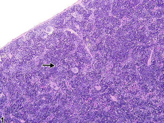
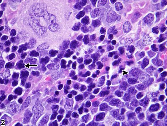
髓外造血(extramedullary hematopoiesis,EMH)在啮齿类动物中常见,是脾红髓的正常组成部分。其在年轻动物中比在老年动物中更常见,在雌性中比在雄性中更常见,在小鼠中比在大鼠中更常见。由于多种情况,如血液毒性损伤、全身性贫血、感染、出血和肿瘤形成,造血细胞数量可能增加至超过正常背景水平(Figure 1、Figure 2、Figure 3、Figure 4)。EMH可包括红系前体细胞(Figure 2、Figure 4,箭头)、髓系前体细胞(Figure 2、Figure 4,箭头head)、巨核细胞(Figure 2、Figure 4)数量增加,或所有三种细胞类型的各种组合。红系前体细胞可继发于出血或红细胞破坏(即溶血性贫血或自身免疫介导的贫血)而占优势,而髓系前体细胞可继发于炎症性、肿瘤性或免疫介导的情况而占优势。浆细胞增生可伴随红髓内的EMH。EMH通常通过其分布和显示不同程度正常分化的造血细胞类型的混合而与肿瘤形成相区分;然而,严重的髓系细胞增殖在组织学上可能类似于粒细胞性白血病。既往术语包括“造血细胞增殖”、“髓系增生”和“红系增生”。
诊断与分级建议(NTP 共识)
仅当髓外造血相对于同期对照的正常背景水平增加或减少(尽管后者罕见)时,才对其进行诊断和分级。髓外造血的诊断应修饰为增加或减少(即,脾 - 髓外造血,增加 或 脾 - 髓外造血,减少)。需要与同期对照进行仔细比较。如果一种细胞类型占优势(即髓系、红系、巨核系),可在病理学叙述中讨论。如果EMH继发于肿瘤形成而增加,则无需记录,但应在病理学叙述中描述。相关链接:肾上腺 - 髓外造血;肝脏 - 髓外造血;淋巴结 - 髓外造血
图谱
英文原文
展开 NTP 官方英文描述与建议
Comment:
Extramedullary hematopoiesis (EMH) is commonly observed in rodents as a normal component of the splenic red pulp. It occurs more frequently in young than in aged animals, in females than in males, and in mice than in rats. Hematopoietic cell numbers may increase above normal background ( Figure 1 , Figure 2 , Figure 3 , and Figure 4 ) due to a variety of conditions, such as hematotoxic insult, systemic anemia, infection, hemorrhage, and neoplasia. EMH may include increased numbers of erythroid precursors ( Figure 2 and Figure 4 , arrows), myeloid precursors ( Figure 2 and Figure 4 , arrowheads), megakaryocytes ( Figure 2 and Figure 4 ), or various combinations of all three cell types. Erythroid precursor cells may predominate secondary to hemorrhage or erythrocyte destruction (i.e., hemolytic anemia or autoimmune-mediated anemia), whereas myeloid precursor cells may predominate secondary to inflammatory, neoplastic, or immune-mediated conditions. Plasma cell hyperplasia may accompany EMH within the red pulp. EMH is generally distinguished from neoplasia by its distribution and mixture of hematopoietic cell types showing various degrees of normal differentiation; however, severe myeloid cell proliferation may resemble granulocytic leukemia histologically. Previous terms include "hematopoietic cell proliferation," "myeloid hyperplasia," and "erythroid hyperplasia."
Recommendation:
Diagnose and grade extramedullary hematopoiesis only when it is increased or decreased (though this is rare) relative to the normal background level in the concurrent controls. The diagnosis of extramedullary hematopoiesis should be modified as increased or decreased (i.e., Spleen - Extramedullary hematopoiesis, Increased or Spleen - Extramedullary hematopoiesis, Decreased). Careful comparison with concurrent controls is necessary. If one cell type predominates (i.e., myeloid, erythroid, megakaryocytic), this can be discussed in the pathology narrative. If EMH is increased secondary to neoplasia, it need not be recorded but should be described in the pathology narrative. Related Links: Adrenal Gland - Extramedullary Hematopoiesis Liver - Extramedullary Hematopoiesis Lymph Node - Extramedullary Hematopoiesis
来源与引用
原始条目:https://ntp.niehs.nih.gov/atlas/nnl/immune-system/spleen/ExtramedullaryHematopoiesis
图谱总目录:NTP Nonneoplastic Lesion Atlas。 内容属美国联邦政府作品(公有领域),可引用;引用时请标注来源。
建议引用格式:NTP Nonneoplastic Lesion Atlas. Extramedullary Hematopoiesis (spleen). LabSmart 中文版:
https://www.labsmart.cn/ntp/spleen-extramedullary-hematopoiesis.html
中文图谱总页(可展开对照全部 595 条):https://www.labsmart.cn/ntp-atlas.html#spleen-extramedullary-hematopoiesis