纤维化Fibrosis · 脾脏 · 免疫系统 · NTP ntp-0040
诊断要点:脾脏纤维化无论何时出现,均应作出诊断并赋予严重程度分级。诊断中应包含部位修饰词(即parenchyma或capsule),以指明纤维化的位置。
病变描述
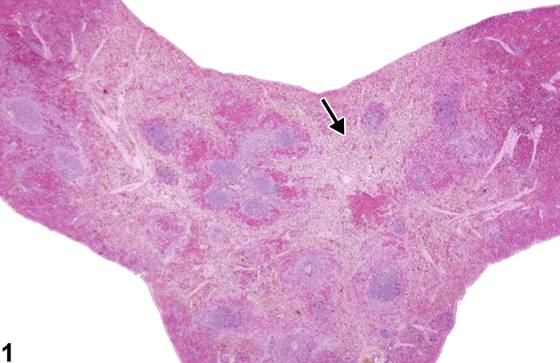
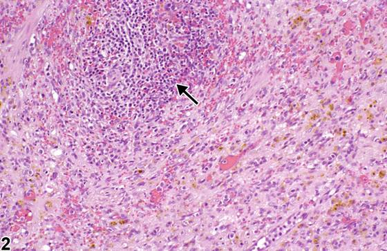
纤维化(fibrosis)的特征是脾脏内胶原性间质沉积增加。纤维化可作为损伤(创伤或毒性)、炎症、梗死或肿瘤形成后的修复过程出现。它可能局限于被膜,也可能作为局灶性(focal)或弥漫性(diffuse)病变发生于实质和/或被膜下区域。实质纤维化可延伸至边缘区并环绕或浸润动脉周围淋巴鞘(Figure 1,箭头);当病变为显著(marked)时,淋巴组织常萎缩(Figure 2,箭头)。被膜纤维化更为常见,通常表现为localized病变(Figure 3、Figure 4,箭头),尽管它可与实质纤维化同时发生。被膜和/或实质纤维化区域内可能存在出血和/或色素,如含铁血黄素和蜡样脂褐素(Figure 2)。红髓的脾窦可能变窄并含少量红细胞。血管可能扩张(angiectasis)。广泛纤维化可导致脾脏塌陷或皱缩(Figure 1)。被膜纤维化可伴有间皮细胞增生或肥大(Figure 4)。纤维化在B6C3F1 mouse中是不常见的背景病变,但已在未处理的大鼠中检出。化学诱导的纤维化可能导致肉瘤形成。
诊断与分级建议(NTP 共识)
脾脏纤维化无论何时出现,均应作出诊断并赋予严重程度分级。诊断中应包含部位修饰词(即parenchyma或capsule),以指明纤维化的位置。被认为继发于脾脏肿瘤形成或炎症的脾纤维化不应单独诊断,除非严重程度足以支持单独诊断,但应在病理学叙述中予以描述。
图谱
英文原文
展开 NTP 官方英文描述与建议
Comment:
Fibrosis is characterized by increased deposition of collagenous stroma in the spleen. Fibrosis can occur as a reparative process following injury (trauma or toxicity), inflammation, infarction, or neoplasia. It may be limited to the capsule or occur as a focal or diffuse lesion in the parenchyma and/or subcapsular region. Parenchymal fibrosis can extend into the marginal zone and surround or infiltrate the periarteriolar lymphatic sheaths ( Figure 1 , arrow); when marked, lymphoid tissue is often atrophied ( Figure 2 , arrow). Capsular fibrosis is more common and typically occurs as a localized lesion ( Figure 3 and Figure 4 , arrows), although it can occur concurrently with parenchymal fibrosis. Hemorrhage and/or pigments such as hemosiderin and ceroid/lipofuscin may be present within regions of capsular and/or parenchyma fibrosis ( Figure 2 ). Sinusoids of the red pulp may be narrowed and contain few erythrocytes. Blood vessels may become ectatic (angiectasis). Extensive fibrosis may result in a collapsed or contracted spleen ( Figure 1 ). Mesothelial hyperplasia or hypertrophy may accompany capsular fibrosis ( Figure 4 ). Fibrosis is an uncommon background lesion in the B6C3F1 mouse, but it has been identified in untreated rats. Chemically induced fibrosis may lead to sarcoma formation.
Recommendation:
Whenever present, fibrosis of the spleen should be diagnosed and assigned a severity grade. A site modifier (i.e., parenchyma or capsule) should be included in the diagnosis to indicate the location of the fibrosis. Splenic fibrosis that is considered to be secondary to neoplasia or inflammation in the spleen should not be diagnosed separately unless warranted by severity, but should be described in the pathology narrative.
来源与引用
原始条目:https://ntp.niehs.nih.gov/atlas/nnl/immune-system/spleen/Fibrosis
图谱总目录:NTP Nonneoplastic Lesion Atlas。 内容属美国联邦政府作品(公有领域),可引用;引用时请标注来源。
建议引用格式:NTP Nonneoplastic Lesion Atlas. Fibrosis (spleen). LabSmart 中文版:
https://www.labsmart.cn/ntp/spleen-fibrosis.html
中文图谱总页(可展开对照全部 595 条):https://www.labsmart.cn/ntp-atlas.html#spleen-fibrosis