淋巴细胞增生Hyperplasia Lymphocyte · 脾脏 · 免疫系统 · NTP ntp-0036
诊断要点:脾脏淋巴细胞增生无论何时出现,均应作出诊断并赋予严重程度分级。脾脏中淋巴细胞的数量通常存在变异;
病变描述
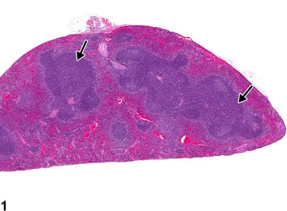
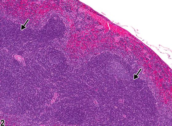
脾脏淋巴细胞增生是指淋巴细胞数量超出同期对照的正常范围。其结果是白髓成分(动脉周围淋巴鞘[PALS]、淋巴滤泡和/或边缘区)的面积和数量出现不同程度的增加(Figure 1、Figure 2,箭头)。该病变可为局灶性(focal)或generalized。脾脏淋巴细胞增生在小鼠中比在大鼠中更常见,且雌性小鼠比雄性小鼠更常受累。显著(marked)淋巴细胞增生可能难以与脾脏早期淋巴瘤区分;然而,淋巴细胞增生时脾脏结构(白髓和红髓分区)应保持完整(Figure 1、Figure 2),而淋巴瘤则预期会出现脾脏结构的丧失。在generalized淋巴细胞增生时,PALS区域可能出现融合(Figure 1),因此应评估细胞形态以与淋巴瘤鉴别。淋巴瘤由单调(或多形性)的肿瘤性淋巴细胞组成,细胞异型性增加,而淋巴细胞增生则由数量增多的正常、成熟淋巴细胞组成。
诊断与分级建议(NTP 共识)
脾脏淋巴细胞增生无论何时出现,均应作出诊断并赋予严重程度分级。脾脏中淋巴细胞的数量通常存在变异;因此,病理学家必须仔细检查并通过与同期对照比较来确定某一研究的正常范围。评估脾脏时还应考虑切面方向(横切面、纵切面、切线切面或浅切面)。诊断应基于病理学家的最佳判断。
图谱
英文原文
展开 NTP 官方英文描述与建议
Comment:
Lymphocyte hyperplasia of the spleen is an increase in the number of lymphocytes outside the range of normal compared with concurrent controls. The result can be variable increases in the area and number of white pulp elements (periarteriolar lymphoid sheaths [PALS], follicles, and/or marginal zones) ( Figure 1 and Figure 2 , arrows). This lesion can be focal or generalized. Lymphocyte hyperplasia in the spleen occurs more frequently in mice than in rats, and female mice are more commonly affected than male mice. Marked lymphocyte hyperplasia can be difficult to distinguish from early lymphoma of the spleen; however, the splenic architecture (white pulp and red pulp compartments) should be maintained with lymphocyte hyperplasia ( Figure 1 and Figure 2 ), whereas loss of the splenic architecture would be expected with lymphoma. With generalized lymphocyte hyperplasia, there can be confluency of the PALS regions ( Figure 1 ), so cellular morphology should be evaluated to differentiate from lymphoma. Lymphoma consists of monomorphic (or pleomorphic) neoplastic lymphocytes with increased cellular atypia, whereas lymphocyte hyperplasia consists of increased numbers of normal, mature lymphocytes.
Recommendation:
Whenever present, lymphocyte hyperplasia of the spleen should be diagnosed and assigned a severity grade. The number of lymphocytes in the spleen is often variable; therefore, the pathologist must carefully examine and determine the range of normal for a study by comparing with concurrent controls. The plane of section should also be taken into consideration (transverse, longitudinal, tangential, or shallow sections) when evaluating the spleen. Diagnosis should be based upon the pathologist’s best judgment.
来源与引用
原始条目:https://ntp.niehs.nih.gov/atlas/nnl/immune-system/spleen/HyperplasiaLymphocyte
图谱总目录:NTP Nonneoplastic Lesion Atlas。 内容属美国联邦政府作品(公有领域),可引用;引用时请标注来源。
建议引用格式:NTP Nonneoplastic Lesion Atlas. Hyperplasia Lymphocyte (spleen). LabSmart 中文版:
https://www.labsmart.cn/ntp/spleen-hyperplasia-lymphocyte.html
中文图谱总页(可展开对照全部 595 条):https://www.labsmart.cn/ntp-atlas.html#spleen-hyperplasia-lymphocyte