肥大细胞增生Hyperplasia Mast Cell · 脾脏 · 免疫系统 · NTP ntp-0041
诊断要点:脾脏肥大细胞增生无论何时出现,均应作出诊断并赋予严重程度分级。
病变描述
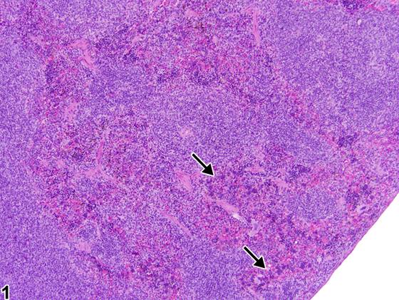
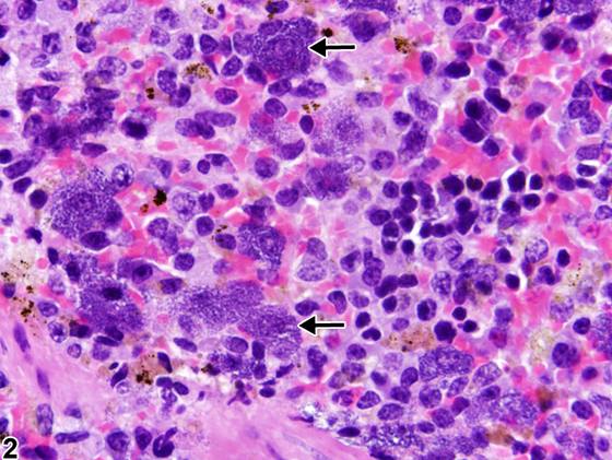
在啮齿类动物的脾脏中可见肥大细胞增生(mast cell hyperplasia)。该病变的特征是脾红髓内出现分化良好的肥大细胞疏松聚集,且不与炎症相关。肥大细胞是相对较大的细胞,胞质丰富,通常含有大量密集的嗜碱性胞质内颗粒,这些颗粒可能遮盖细胞核(Figure 1、Figure 2,箭头),但也可能表现为明显的淡嗜碱性胞质和不太明显的颗粒。肥大细胞增生必须与转移性肥大细胞瘤相鉴别。在无原发性肥大细胞瘤的情况下,脾脏内出现肥大细胞支持增生性病变而非肿瘤性病变。
诊断与分级建议(NTP 共识)
脾脏肥大细胞增生无论何时出现,均应作出诊断并赋予严重程度分级。
图谱
英文原文
展开 NTP 官方英文描述与建议
Comment:
Mast cell hyperplasia can be seen within the spleen of rodents. This lesion is characterized by loose aggregates of well-differentiated mast cells within the splenic red pulp that are not associated with inflammation. Mast cells are relatively large cells with abundant cytoplasm and typically have numerous densely basophilic intracytoplasmic granules that may obscure the nucleus ( Figure 1 and Figure 2 , arrows), although they may also present with prominent pale basophilic cytoplasm and less apparent granules. Mast cell hyperplasia must be differentiated from metastatic mast cell tumors. Mast cells within the spleen in the absence of a primary mast cell tumor are supportive of a hyperplastic rather than a neoplastic lesion.
Recommendation:
Whenever present, mast cell hyperplasia of the spleen should be diagnosed and assigned a severity grade.
来源与引用
原始条目:https://ntp.niehs.nih.gov/atlas/nnl/immune-system/spleen/HyperplasiaMastCell
图谱总目录:NTP Nonneoplastic Lesion Atlas。 内容属美国联邦政府作品(公有领域),可引用;引用时请标注来源。
建议引用格式:NTP Nonneoplastic Lesion Atlas. Hyperplasia Mast Cell (spleen). LabSmart 中文版:
https://www.labsmart.cn/ntp/spleen-hyperplasia-mast-cell.html
中文图谱总页(可展开对照全部 595 条):https://www.labsmart.cn/ntp-atlas.html#spleen-hyperplasia-mast-cell