色素Pigment · 脾脏 · 免疫系统 · NTP ntp-0043
诊断要点:当脾红髓和/或白髓中的色素因受试物而增多或减少时,应作出诊断并赋予严重程度分级。
病变描述
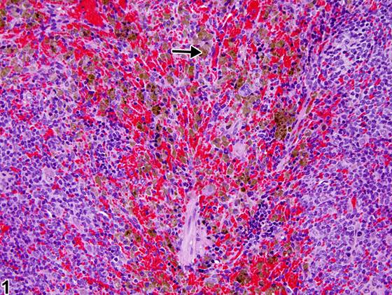
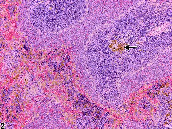
脾红髓巨噬细胞内存在少量胞质内色素是啮齿类动物的常见背景性所见。色素可为含铁血黄素(hemosiderin)、蜡样质/脂褐素(ceroid/lipofuscin)、黑色素(melanin)或受试物相关色素。色素通常见于红髓(Figure 1,箭头),但也可见于白髓的边缘区和/或动脉周围淋巴鞘(Figure 2,箭头)、被膜或小梁。含铁血黄素色素是巨噬细胞吞噬红细胞(通常为衰老或受损红细胞)或游离血红蛋白的结果,在老龄啮齿类动物和/或治疗诱导的溶血性贫血或高铁血红蛋白血症、慢性充血、造血细胞增殖增加以及红细胞吞噬作用等情况下可能增多。含铁血黄素沉积也可见于继发于恶性肿瘤的局灶性(focal)出血。在老龄啮齿类动物中,大鼠和小鼠含铁血黄素色素的发生率通常均为雌性高于雄性。蜡样质/脂褐素来源于不饱和脂质的氧化和聚合,也可存在于脾巨噬细胞内,但较含铁血黄素少见。含铁血黄素可用Perl’s普鲁士蓝特殊染色识别,而蜡样质/脂褐素呈抗酸染色阳性。黑色素色素可见于小梁黑色素细胞或黑色小鼠红髓的局灶性区域。Fontana-Masson染色或Schmorl’s铁氰化铁还原染色可用于识别黑色素颗粒。只有当特殊染色证实后,色素才可按类型(即含铁血黄素、蜡样质/脂褐素、黑色素)进行亚分类。
诊断与分级建议(NTP 共识)
当脾红髓和/或白髓中的色素因受试物而增多或减少时,应作出诊断并赋予严重程度分级。诊断中应包含诊断修饰词(即“increased”或“decreased”),以表明色素相对于同期对照是增多还是减少。即使使用一系列特殊染色,在组织学切片中明确鉴定色素类型通常仍很困难。因此,最恰当的诊断是“色素”(而非诊断色素类型,例如含铁血黄素或脂褐素)。色素的形态学特征应在病理学叙述中描述。如果色素的存在继发于年龄或无毒理学意义,则不应作出色素诊断。病理学家应运用其判断来决定继发性色素沉积是否显著到足以需要单独诊断。
图谱
英文原文
展开 NTP 官方英文描述与建议
Comment:
The presence of a small amount of intracytoplasmic pigment within the splenic red pulp macrophages is a common background finding in rodents. Pigment can be hemosiderin, ceroid/lipofuscin, melanin, or test article related. It is typically found in the red pulp ( Figure 1 , arrow) but may also be present in the marginal zone and/or periarteriolar lymphatic sheaths of the white pulp ( Figure 2 , arrow), the capsule, or trabeculae. Hemosiderin pigment is the result of macrophage engulfment of red blood cells (usually effete or damaged) or free hemoglobin and may be increased in aging rodents and/or such conditions as treatment-induced hemolytic anemia or methemoglobinemia, chronic congestion, increased hematopoietic cell proliferation, and erythrophagocytosis. Hemosiderin deposits may also be observed with focal hemorrhage secondary to malignant neoplasms. The incidence of hemosiderin pigment in aging rodents is usually higher in females than in males for both rats and mice. Ceroid/lipofuscin, derived from oxidation and polymerization of unsaturated lipids, may also be present within splenic macrophages but occurs less commonly than hemosiderin. Hemosiderin can be identified with Perl’s Prussian blue special stain, whereas ceroid/lipofuscin is acid-fast positive. Melanin pigment can be seen in trabecular melanocytes or focally in the red pulp of black mice. Fontana-Masson or Schmorl’s ferric-ferricyanide reduction stain can be used to identify melanin granules. Pigment may be subclassified by type (i.e., hemosiderin, ceroid/ lipofuscin, melanin) only when confirmed by special stains.
Recommendation:
Pigment in the red and/or white pulp of the spleen should be diagnosed when increased or decreased due to treatment and assigned a severity grade. A diagnostic modifier (i.e., "increased" or "decreased") should be included in the diagnosis to indicate whether the pigment is increased or decreased relative to concurrent controls. Definitive pigment identification is often difficult in histological sections, even with a battery of special stains. Therefore, a diagnosis of "pigment" (as opposed to diagnosing the type of pigment, e.g., hemosiderin or lipofuscin) is most appropriate. Morphological features of the pigment should be described in the pathology narrative. If the presence of pigment is secondary to age or not toxicologically meaningful, the pigment should not be diagnosed. The pathologist should use his or her judgment in deciding whether secondary deposits of pigment are prominent enough to warrant a separate diagnosis.
来源与引用
原始条目:https://ntp.niehs.nih.gov/atlas/nnl/immune-system/spleen/Pigment
图谱总目录:NTP Nonneoplastic Lesion Atlas。 内容属美国联邦政府作品(公有领域),可引用;引用时请标注来源。
建议引用格式:NTP Nonneoplastic Lesion Atlas. Pigment (spleen). LabSmart 中文版:
https://www.labsmart.cn/ntp/spleen-pigment.html
中文图谱总页(可展开对照全部 595 条):https://www.labsmart.cn/ntp-atlas.html#spleen-pigment