上皮变性Epithelium Degeneration · 前胃 · 消化系统 · NTP ntp-0202
诊断要点:当变性作为原发性病变存在时,应当诊断并分级。当变性作为继发性病变存在时(例如继发于炎症时),不应诊断,但应在病理学描述中予以描述。
病变描述
细胞质空泡化(cytoplasmic vacuolization)、囊性变性(cystic degeneration)和水疱/大疱形成(vesicle/bulla formation)是鳞状上皮变性的几种形式,可发生于体内任何鳞状上皮。鳞状上皮的空泡化表现为表面上皮细胞染色浅淡、细胞质空泡化。相关黏膜可能发生溃疡,上皮细胞可能变性或坏死。下方组织有不同程度的炎症细胞浸润。此变化可能代表较坏死更轻微的改变,可见于采用灌胃方法的研究中。此病变也可能由致溃疡性或抗炎性化合物引起。囊性变性的特征为上皮细胞空泡化和气球样肿胀,导致细胞膜破裂,在上皮内形成不规则间隙,可能进而形成水疱或大疱。相关黏膜可能发生溃疡,上皮细胞可能变性或坏死。下方组织有不同程度的炎症细胞浸润。此变化可能代表空泡变性的进展,可见于采用灌胃方法的研究中。此病变也可能由致溃疡性或抗炎性化合物引起。大疱的特征为上皮层内或上皮层下局限性的液体积聚。水疱<1 cm,大疱≥1.0 cm。大疱可由棘层松解(acantholysis)、表皮或真皮水肿、基底细胞和角质形成细胞变性,或其他过程(如摩擦性创伤和烧伤导致细胞间黏附丧失)所形成。大疱可导致糜烂和溃疡。
诊断与分级建议(NTP 共识)
当变性作为原发性病变存在时,应当诊断并分级。当变性作为继发性病变存在时(例如继发于炎症时),不应诊断,但应在病理学描述中予以描述。如果变性是病变的显著特征但不被视为原发性病变,若病理学家认为有正当理由,可将其与原发性病变同时诊断。分级应基于细胞肿胀的程度以及受累区域的范围和数量。如果变性和坏死同时存在,两者均应诊断并分级。炎症不应单独诊断,除非它是病变的显著特征。
图谱
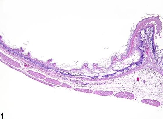
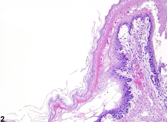
英文原文
展开 NTP 官方英文描述与建议
Comment:
Cytoplasmic vacuolization, cystic degeneration, and vesicle/bulla formation are forms of degeneration of the squamous epithelium that can occur in any squamous epithelium in the body. Vacuolization of the squamous epithelium is characterized by superficial epithelial cells with a pale staining and vacuolated cytoplasm. Associated mucosa may be ulcerated, and epithelial cells may be degenerate or necrotic. Subjacent tissue is variably infiltrated by inflammatory cells. This change may represent a milder change than necrosis and can be observed in studies involving gavage methods. This lesion may also be due to ulcerogenic or anti-inflammatory compounds. Cystic degeneration is characterized by vacuolization and ballooning swelling of epithelial cells, resulting in rupture of cell membranes to form irregular spaces in the epithelium, potentially leading to development of vesicles or bulla. Associated mucosa may be ulcerated, and epithelial cells may be degenerate or necrotic. Subjacent tissue is variably infiltrated by inflammatory cells. This change may represent a progression of vacuolar degeneration and can be observed in studies involving gavage methods. This lesion may also be due to ulcerogenic or anti-inflammatory compounds. Bulla are characterized as a localized collection of fluid in or beneath the epithelial layer. Vesicles are <1 cm and bulla are ≥ 1.0 cm. Bulla can form as the result of acantholysis, epidermal or dermal edema, degeneration of basal cells and keratinocytes, or other processes, such as frictional trauma and burns that cause a lack of cohesion between cells. Bulla can lead to erosions and ulceration.
Recommendation:
Degeneration should be diagnosed and graded when present as a primary lesion. When degeneration is present as a secondary lesion (e.g., when secondary to inflammation) it should not be diagnosed but should be described in the pathology narrative. If the degeneration is a prominent feature of the lesion but is not considered the primary lesion, it may be diagnosed concurrently the primary lesion if the pathologist feels it is warranted. Grading should be based on the degree of cellular swelling and extent and number of areas affected. If both degeneration and necrosis are present, both should be diagnosed and graded. Inflammation should not be diagnosed separately, unless it is a prominent feature of the lesion.
来源与引用
原始条目:https://ntp.niehs.nih.gov/atlas/nnl/alimentary-system/stomach-forestomach/Epithelium-Degeneration
图谱总目录:NTP Nonneoplastic Lesion Atlas。 内容属美国联邦政府作品(公有领域),可引用;引用时请标注来源。
建议引用格式:NTP Nonneoplastic Lesion Atlas. Epithelium Degeneration (stomach-forestomach). LabSmart 中文版:
https://www.labsmart.cn/ntp/stomach-forestomach-epithelium-degeneration.html
中文图谱总页(可展开对照全部 595 条):https://www.labsmart.cn/ntp-atlas.html#stomach-forestomach-epithelium-degeneration