坏死Necrosis · 前胃 · 消化系统 · NTP ntp-0207
诊断要点:如果坏死上皮仍然存在且至少部分附着于其下的固有层,或者坏死并非特别以erv前胃上皮为中心(即主要累及胃壁),则应诊断上皮坏死,而不是糜烂和溃疡。
病变描述
坏死的特征性表现为核固缩(pyknosis)、核碎裂(karyorrhexis)、核溶解(karyolysis)等核改变,或核溶解后期细胞核消失。早期坏死时,H&E切片中细胞质呈均质粉红色。细胞质蛋白降解最终使坏死细胞呈苍白、鬼影状外观。坏死细胞通常失去与基底膜及邻近细胞的黏附。细胞破裂、细胞完整性丧失是细胞死亡最明显的证据。除染毒大鼠外,前胃黏膜坏死罕见。如Figure 1、Figure 2、Figure 3、Figure 4所示,小鼠和大鼠前胃的化学相关病变包括坏死和溃疡。在大鼠中,前胃坏死在灌胃研究中比在掺食或吸入研究中更常见。血栓形成可导致胃坏死(梗死)(Figure 4,箭头)。
诊断与分级建议(NTP 共识)
如果坏死上皮仍然存在且至少部分附着于其下的固有层,或者坏死并非特别以erv前胃上皮为中心(即主要累及胃壁),则应诊断上皮坏死,而不是糜烂和溃疡。如果坏死符合继发于血栓形成的缺血(梗死),则叙述中应说明这一点,即使血栓不在切片平面内。严重程度分级取决于病变的总体大小和受累区域的数量。虽然出血、水肿、纤维化或炎症可能与坏死相关,但这些通常不单独记录,但可在叙述中描述。如果它们是坏死病变的显著组成部分,则应单独诊断。
图谱
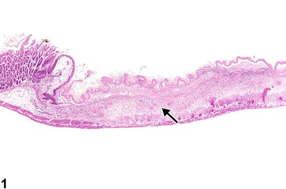
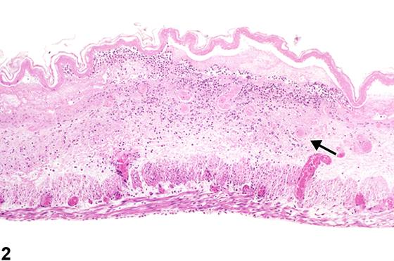
英文原文
展开 NTP 官方英文描述与建议
Comment:
Necrosis is characterized by nuclear changes of pyknosis, karyorrhexis, karyolysis, or absence of nucleus in the later stage of karyolysis. The cytoplasm in early necrosis is homogenous pink in H&E sections. Degradation of cytoplasmic proteins eventually gives the necrotic cell a pale, ghostlike appearance. Necrotic cells usually lose their adherence to basement membranes and neighboring cells. Rupture of cells with loss of cell integrity is the most obvious evidence of cell death. Necrosis of the forestomach mucosa is rarely seen except in treated rats. As shown in Figure 1 , Figure 2 , Figure 3 , and Figure 4 , chemical-related lesions in the forestomach of mice and rats include necrosis and ulceration. Necrosis of the forestomach is more frequent in gavage studies than in dosed-feed or inhalation studies in rats. Thrombosis can result in necrosis (infarction) of the stomach ( Figure 4 , arrow).
Recommendation:
Necrosis of epithelium is diagnosed instead of erosion and ulcer if the necrotic epithelium is still present and at least partially attached to the underlying lamina propria or if the necrosis is not particularly centered on the epithelium of the forestomach (i.e., involves the wall primarily). If the necrosis is consistent with ischemia secondary to thrombosis (infarction), the narrative should state this, even if the thrombus is not in the plane of the section. Severity grading would depend on the overall size of the lesion and number of areas affected. While hemorrhage, edema, fibrosis, or inflammation may be associated with necrosis, these are usually not recorded separately but can be described in the narrative. If they are a prominent component of a necrotic lesion, they should be diagnosed separately.
来源与引用
原始条目:https://ntp.niehs.nih.gov/atlas/nnl/alimentary-system/stomach-forestomach/Necrosis
图谱总目录:NTP Nonneoplastic Lesion Atlas。 内容属美国联邦政府作品(公有领域),可引用;引用时请标注来源。
建议引用格式:NTP Nonneoplastic Lesion Atlas. Necrosis (stomach-forestomach). LabSmart 中文版:
https://www.labsmart.cn/ntp/stomach-forestomach-necrosis.html
中文图谱总页(可展开对照全部 595 条):https://www.labsmart.cn/ntp-atlas.html#stomach-forestomach-necrosis