上皮增生(局灶性/弥漫性)Epithelium Hyperplasia[Focal Diffuse] · 前胃 · 消化系统 · NTP ntp-0203
诊断要点:增生一经出现即应作出诊断并分级。应尽可能使用修饰词“局灶性(focal)”和“弥漫性(diffuse)”。
病变描述
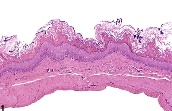
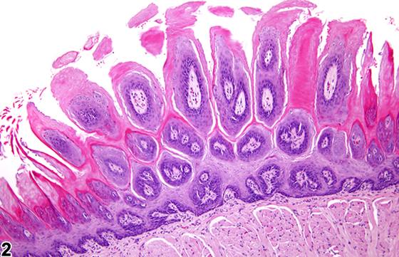
增生(hyperplasia)被定义为上皮一层或多层(基底层、棘层或颗粒层)中细胞数量的增加。当黏膜存在增生时,常可见角化过度(hyperkeratosis,即角质层厚度增加)。增生可为弥漫性(弥漫性(diffuse))(Figure 1、Figure 6、Figure 7、Figure 8)或局灶性(局灶性(focal))(Figure 2、Figure 3、Figure 4),并导致非腺性胃黏膜厚度增加。增生也可见于界嵴(limiting ridge)(Figure 6;与Figure 5中的正常界嵴相比较)。非腺性胃的增生通常表现为上皮各层均匀增厚,而无向上或向下的突起。然而,在更严重的病例中,上皮出现卷曲,并伴有网状钉突样结构(rete peg-like structures)的向下突起。基底细胞可能发生增殖,并可延伸至黏膜下层(Figure 7、Figure 8)。在局灶性增生较严重的病例中,可能出现向管腔内向上增殖、类似乳头状瘤(papilloma)的病变,在描述中可称为乳头状增生(papillary hyperplasia)。缺乏含血管的分支状结缔组织核心以及黏膜肌层完整,可将这种形式的增生与真性乳头状瘤区分开来。增生时,仍保留一定程度的有序成熟。
诊断与分级建议(NTP 共识)
增生一经出现即应作出诊断并分级。应尽可能使用修饰词“局灶性(focal)”和“弥漫性(diffuse)”。分级应基于病变大小、分布范围及上皮厚度。多灶性病变不记录为“多灶性(multifocal)”,但应给予比单发病变更高的严重程度分级。若存在异型性特征,如角化不良(diskeratinization)、结构紊乱(disorganization)或存在非典型细胞,则应诊断为非典型增生(非典型性(atypical) hyperplasia)(见 Stomach, Forestomach - Epithelium - Hyperplasia, 非典型性)。若病理学家认为存在两种不同的增生过程(例如弥漫性和非典型性),则两者均应作出诊断,并在病理学描述中详尽描述。与增生相关的角化过度通常不作为独立病变诊断,除非它是病变的显著组成部分,但应在病理学描述中予以描述。相关链接:Stomach, Forestomach - Epithelium - Hyperplasia, 非典型性
图谱
英文原文
展开 NTP 官方英文描述与建议
Comment:
Hyperplasia is defined as an increase in the number of cells in one or more epithelial strata (basal, spinous, or granulosum). Hyperkeratosis (increased thickness of the stratum corneum) is often seen when hyperplasia of the mucosa is present. Hyperplasia may be diffuse ( Figure 1 , Figure 6 , Figure 7 , and Figure 8 ) or focal ( Figure 2 , Figure 3 , and Figure 4 ) and results in an increased thickness of the non-glandular stomach mucosa. Hyperplasia may also be seen at the limiting ridge ( Figure 6 ; compare with normal limiting ridge in Figure 5 ). Hyperplasia of the nonglandular stomach is often characterized as a uniform thickening of all layers of the epithelium without upward or downward projections. However, in more severe cases there are convolutions of the epithelium with downward projections of rete peg-like structures. There may be proliferation of the basal cells, which can extend into the submucosa ( Figure 7 and Figure 8 ). Focal upward proliferation into the lumen mimicking papilloma may be present in more severe cases of focal hyperplasia and may be described as papillary hyperplasia in the narrative. The absence of a branching connective tissue core containing blood vessels and the presence of an intact muscularis mucosa distinguish this form of hyperplasia from a true papilloma. With hyperplasia, there remains some degree of orderly maturation.
Recommendation:
Hyperplasia should be diagnosed and graded whenever present. The modifiers "focal" and "diffuse" should be used whenever possible. Grading should be based on the size, extent of distribution, and thickness of the epithelium. Multifocal lesions are not recorded as "multifocal" but are given a higher severity grade than single lesions. If there are features of atypia, such as diskeratinization, disorganization, or the presence of atypical cells, then atypical hyperplasia should be diagnosed (see Stomach, Forestomach - Epithelium - Hyperplasia, Atypical ). If the pathologist feels that two distinct hyperplastic processes are present (e.g., diffuse and atypical), then both should be diagnosed and thoroughly described in the pathology narrative. Hyperkeratosis associated with hyperplasia is usually not diagnosed as a separate entity, unless it is a prominent component of the lesion, but it should be described in the pathology narrative. Related Links: Stomach, Forestomach - Epithelium - Hyperplasia, Atypical
来源与引用
图谱总目录:NTP Nonneoplastic Lesion Atlas。 内容属美国联邦政府作品(公有领域),可引用;引用时请标注来源。
建议引用格式:NTP Nonneoplastic Lesion Atlas. Epithelium Hyperplasia[Focal Diffuse] (stomach-forestomach). LabSmart 中文版:
https://www.labsmart.cn/ntp/stomach-forestomach-epithelium-hyperplasia-focal-diffuse.html
中文图谱总页(可展开对照全部 595 条):https://www.labsmart.cn/ntp-atlas.html#stomach-forestomach-epithelium-hyperplasia-focal-diffuse