滤泡细胞肥大Follicular Cell Hypertrophy · 甲状腺 · 内分泌系统 · NTP ntp-0066
诊断要点:甲状腺滤泡细胞肥大存在时应当诊断并分级。病理学描述可说明肥大滤泡上皮的性质,以及是否为双侧、是否伴有滤泡增生,或是否与胶质改变相关。
病变描述
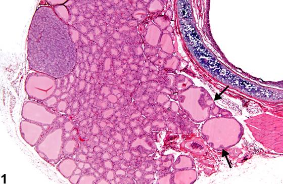
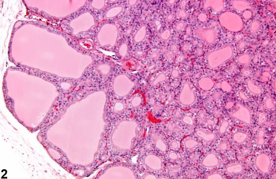
在滤泡细胞肥大(follicular cell hypertrophy)中,甲状腺滤泡可能由立方状至柱状上皮衬覆,中央滤泡紧密排列且小于正常(Figure 1、Figure 2)。滤泡上皮的局灶性(focal)增生有时可伴随滤泡肥大(Figure 1,箭头)。双侧肥大是对垂体促甲状腺激素分泌增加的系统性反应,属预期所见。肥大的滤泡上皮可含有淡嗜酸性胞质和小而清亮的空泡(Figure 3)。在更为慢性的滤泡上皮肥大中,所有滤泡均可能小于正常,伴有分泌耗竭或分泌异常及上皮脱落(Figure 4)。甲状腺滤泡细胞肥大的出现可能继发于肝细胞微粒体酶诱导,并且在甲状腺滤泡细胞肥大的大鼠中常同时可见肝细胞肥大。
诊断与分级建议(NTP 共识)
甲状腺滤泡细胞肥大存在时应当诊断并分级。病理学描述可说明肥大滤泡上皮的性质,以及是否为双侧、是否伴有滤泡增生,或是否与胶质改变相关。
图谱
英文原文
展开 NTP 官方英文描述与建议
Comment:
In follicular cell hypertrophy, thyroid follicles may be lined by cuboidal to columnar epithelium with central follicles tightly packed and smaller than normal ( Figure 1 and Figure 2 ). Focal hyperplasia of follicular epithelium may sometimes accompany follicular hypertrophy ( Figure 1 , arrows). Bilateral hypertrophy is expected as a systemic response to increased pituitary secretion of thyroid-stimulating hormone. Hypertrophic follicular epithelium may contain pale eosinophilic cytoplasm and small clear vacuoles ( Figure 3 ). In more chronic follicular epithelial hypertrophy, all follicles may be smaller than normal, with secretory depletion or abnormal secretion and exfoliated epithelium ( Figure 4 ). The presence of thyroid follicular cell hypertrophy may be secondary to hepatocellular microsomal enzyme induction, and hepatocellular hypertrophy is often seen concomitantly in rats with thyroid gland follicular cell hypertrophy.
Recommendation:
Thyroid follicular cell hypertrophy should be diagnosed and graded when present. The pathology narrative can describe the nature of the hypertrophic follicular epithelium and whether bilateral, accompanied by follicular hyperplasia, or associated with alterations in colloid.
来源与引用
原始条目:https://ntp.niehs.nih.gov/atlas/nnl/endocrine-system/thyroid-gland/FollicularCell-Hypertrophy
图谱总目录:NTP Nonneoplastic Lesion Atlas。 内容属美国联邦政府作品(公有领域),可引用;引用时请标注来源。
建议引用格式:NTP Nonneoplastic Lesion Atlas. Follicular Cell Hypertrophy (thyroid-gland). LabSmart 中文版:
https://www.labsmart.cn/ntp/thyroid-gland-follicular-cell-hypertrophy.html
中文图谱总页(可展开对照全部 595 条):https://www.labsmart.cn/ntp-atlas.html#thyroid-gland-follicular-cell-hypertrophy