轴索病Axonopathy · 脑 · 神经系统 · NTP ntp-0398
诊断要点:在NTP研究中,所有形式的轴索损伤均首选“轴索病(axonopathy)”一词。
病变描述
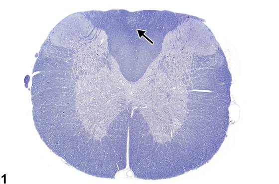
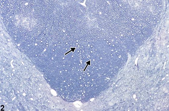
“轴索病(axonopathy)”是一个通用术语,描述发生于脑、脊髓和周围神经的多种轴索病变。在低倍镜下必须注意,确保轴索病的判定标准(轴索肿胀、断裂、丢失等)不被误认为人为空泡化而忽视。其在脑和脊髓特定区域的出现以及常呈对称性分布,是判断其为真实病变的有用线索。需要仔细检查白质和神经组织的所有区域,才能正确发现和解释此类病变。年龄相关轴索病常见于马尾、腹侧脊神经根、脊髓腹索和侧索、坐骨神经和臂神经以及低位脑干。对老龄大鼠脊髓和神经根性(神经根)轴索病的解释应谨慎,因为它可能是衰老的结果,在24月龄或以上大鼠中的发生率为75-90%。在专门的毒理学-神经病理学研究中,使用特殊轴索染色如Bielschowsky银浸染和de Olmos氨基铜银染色分别突出显示正常轴索和轴索病变,可能有助于界定这些常较细微病变的严重程度和分布。Figure 1显示低倍镜下脊髓,展示背侧本体感觉索即本例中薄束内轴索病的表现。注意在此放大倍数下,病变表现为通常由正常上行轴索占据的局灶性(focal)区域出现细微空泡化(箭头)。该区域轴索受损导致轴索及其髓鞘肿胀,从而形成扩张的空隙。此图采用甲苯胺蓝染色。Figure 2为亚慢性毒性研究中脊髓薄束轴索肿胀的甲苯胺蓝染色高倍切片。注意许多膨大的轴索(箭头),大多数仍被其髓鞘包围。有髓纤维明显减少,尤其在索的最深部,使这些区域肿胀的有髓轴索如大“空泡”般凸显。Figure 3、Figure 4、Figure 5描绘肿胀的轴索。在Figure 3中,注意扩张空隙中央偶见轻度增大的肿胀轴索(箭头),该空隙原先由正常、致密的板层状髓鞘占据。许多扩张空隙是空的,因为轴索已经发生崩解、溶解并被巨噬细胞吞噬。空隙看似空虚是因为组织处理中使用的脂肪溶剂去除了构成髓鞘的残留复合脂质。Figure 4代表更高放大倍数下的肿胀退变轴索(箭头),其周围扩张空隙的直径约为相邻外观正常轴索周围空隙的三倍。Figure 5代表脊髓索轴索病,详示一个肿胀轴索,即所谓的球状体(spheroid)(箭头)。注意空泡内一个核固缩的巨噬细胞,代表轴索变性后期阶段,特征为轴索被吞噬(箭头)。在大多数轴索变性病例中,可识别的巨噬细胞通常已坏死,推测是由于轴索变性过程中吞噬髓鞘脂质和释放细胞因子的影响。当巨噬细胞负载游离胆固醇和三酰甘油时,即发生凋亡。在此高倍苏木精-伊红染色图像中,大的肿胀轴索呈双嗜性外观。这是轴索肿胀早期、即轴索崩解之前球状体的良好示例。Figure 6显示脑切片中嗜酸性球状体和卵圆形至长条形结构(箭头),代表受损轴索,因切片平面不同而形态各异。脑内肿胀轴索的出现通常不伴有脊髓和周围神经中所见的髓鞘显著扩张。另请注意该区域混合性胶质细胞群轻度增多,为相关炎症反应的一部分。Figure 7显示轴索变性伴典型“消化腔(digestion chamber)”,内含轴索碎片(箭头)。在此坐骨神经纵切面苏木精-伊红染色图像中,称为沃勒变性(Wallerian degeneration)的轴索变性典型表现清晰可见。“沃勒变性”一词最好保留用于描述周围神经的轴索病;然而,脊髓和脑内也可见类似改变。Augustus Waller于1850年通过对实验性神经挤压伤的序贯研究,提出了周围神经轴索病的判定标准。远端神经尤其在挤压伤后,特征为进行性轴索肿胀,并在挤压后约36小时出现纤维断裂、Schwann细胞核肥大、巨噬细胞浸润以及所谓消化腔的形成。在Figure 7中,消化腔含数个退变轴索碎片,周围为含脂质分解产物和巨噬细胞的透明空隙。相邻神经组织中可见Schwann细胞增大的核。Figure 8显示坐骨神经轴索变性伴Schwann细胞反应性增生和核肥大(箭头)。这些呈线性带状增殖的Schwann细胞称为Büngner带(bands of Büngner)。它们是在轴索损伤后Schwann细胞失去其轴索联系时作为反应而形成的。残留的基底膜形成神经内膜管,对引导近端再生轴索芽生十分重要。Schwann细胞和血源性巨噬细胞在吞噬断裂轴索以清除神经内膜管、利于轴索再生方面起重要作用。Büngner带不应解释为炎性纤维化,而应视为周围轴索显著变性后神经再支配过程的一部分。
诊断与分级建议(NTP 共识)
在NTP研究中,所有形式的轴索损伤均首选“轴索病(axonopathy)”一词。该病变应在对照和治疗动物中诊断,并注明其解剖亚部位。严重程度分级应连同判定标准一并记录,以便根据发生率和严重程度决定其与治疗的相关性。努力寻找轴索病的明确判定标准,对于在病变演变的多个阶段确认其真实性质十分重要。在NTP研究中,若研究病理学家对白质局灶性(focal)空泡性病变的发病机制不确定,或病变无法与脱髓鞘或髓鞘内水肿区分,则该病变应诊断为白质空泡化(white matter vacuolation)。描述应说明病变为局灶性、单侧还是双侧等特征。随后应由神经病理学家进行专门评估。
图谱
英文原文
展开 NTP 官方英文描述与建议
Comment:
“Axonopathy” is a generic term describing a variety of lesions of the axon occurring in brain, spinal cord, and peripheral nerve. Care must be taken at low magnifications to ensure that the criteria for axonopathy (axonal swelling, fragmentation, loss, etc.) are not disregarded as artifactual vacuolation. Its presence in defined regions of brain and spinal cord and frequent symmetrical distribution are useful clues to its genuine nature. Close examination of all regions of white matter and nerve tissue is required to correctly discover and interpret this type of lesion. Age-related axonopathy occurs commonly in the cauda equina, ventral spinal nerve roots, ventral and lateral spinal funiculi, sciatic and brachial nerves, and lower brainstem. Caution should be given to interpretation of spinal and radicular (nerve root) axonopathy in aged rats since it may be a result of senescence and has an incidence of 75-90% in rats 24 or more months of age. In specialized toxicologic-neuropathologic studies, use of special axonal stains such as Bielschowsky silver impregnation and de Olmos amino cupric silver to highlight the normal axon and axonal lesions, respectively, may be helpful in delineating the severity and distribution of these often subtle lesions. Figure 1 shows spinal cord at low magnification demonstrating the appearance of axonopathy in the dorsal proprioceptive funiculi, in this case the fasciculus gracilis. Note, at this magnification, that the lesion is represented by subtle vacuolation (arrow) of a focal area ordinarily populated by normal ascending axons. Damage to the axons in this zone leads to swelling of both the axon and its myelin sheath, thus creating a dilated space. This image uses toluidine blue stain. Figure 2 is a higher-magnification toluidine blue–stained section of spinal cord showing axonal swelling in the fasciculus gracilis in a subchronic toxicity study. Note the many distended axons (arrows), most still surrounded by their myelin sheath. There is a prominent reduction in myelinated fibers, particularly in the deepest parts of the funiculi, making the swollen myelinated axons in these regions stand out in relief as large ”vacuoles.” Figure 3 , Figure 4 , and Figure 5 depict swollen axons. In Figure 3 , note the slightly enlarged occasional swollen axons (arrows) at the center of dilated spaces formerly occupied by a normal, compact lamellar myelin sheath. Many of the dilated spaces are empty because the axon has already undergone disintegration, lysis, and phagocytosis by macrophages. The space appears empty because the use of fat solvents in tissue processing has removed any remaining complex lipids, which made up the myelin sheath. Figure 4 represents swollen degenerate axons at higher magnification (arrows), where the diameter of the distended space surrounding them is approximately three times larger than the space around adjacent, apparently normal axons. Figure 5 represents spinal funicular axonopathy with detail of a swollen axon, a so-called spheroid (arrow). Note the pyknotic macrophage in a vacuole representing a later stage of axonal degeneration characterized by axonal phagocytosis (arrowhead). In most cases of axonal degeneration, identifiable macrophages are usually necrotic, presumably from the effects of myelin lipid phagocytosis and cytokine release during the axonal degenerative process. Apoptosis of macrophages results when they are loaded with free cholesterol and triacylglycerol. In this high-magnification hematoxylin and eosin–stained image, the large swollen axon has an amphophilic appearance. This is a good example of a spheroid at an early stage of axonal swelling that precedes axonal disintegration. Figure 6 shows eosinophilic spheroids and oval to elongated structures (arrows) that represent injured axons in the brain sectioned in various planes, thus affecting their appearance. The presence of swollen axons in the brain is not generally accompanied by dramatic distension of the myelin sheath such as those seen in spinal cord and peripheral nerves. Note also that there is a mild increase in a mixed population of glial cells in the region, part of the associated inflammatory response. Figure 7 shows axon degeneration with a typical “digestion chamber,” containing axonal fragments (arrow). In this hematoxylin and eosin–stained image of a longitudinal section of the sciatic nerve, the typical appearance of axonal degeneration referred to as Wallerian degeneration is evident. The term “Wallerian degeneration” is best reserved to describe axonopathy in peripheral nerve; however, similar changes can be seen in spinal cord and brain. Augustus Waller, in 1850, introduced the criteria for axonopathy in peripheral nerve from his sequential studies of experimental nerve crush injury. The distal nerve, particularly after crush, is characterized by progressive axonal swelling and, approximately 36 hours after crush, by fragmentation of the fiber, Schwann cell nuclear hypertrophy, infiltration of macrophages, and the formation of so-called digestion chambers. In Figure 7 the digestion chamber contains several fragments of degenerate axon surrounded by a clear space, containing lipid breakdown products and macrophages. The enlarged nuclei of Schwann cells can be seen in adjacent nerve tissue. Figure 8 shows sciatic nerve axon degeneration with reactive hyperplasia and hypertrophic nuclei of Schwann cells (arrows). These linear band-like proliferative Schwann cells are known as bands of Büngner. They are formed as a response to axonal injury when Schwann cells lose their axonal complement. The remaining basement membrane forms endoneurial tubes, important in the guidance of regenerating proximal axonal sprouts. Schwann cells and blood-borne macrophages are important in phagocytosis of fragmented axons to clear endoneurial tubes for axonal regeneration. The bands of Büngner should not be interpreted as inflammatory fibrosis but, rather, as a part of the reinnervation process subsequent to significant axon degeneration in peripheral axons.
Recommendation:
In NTP studies, the term “axonopathy” is preferred for all forms of axonal injury. This lesion should be diagnosed in control and treated animals with its anatomic subsite location. Severity grade should be included with criteria, allowing a decision on its treatment-related significance to be based on incidence and severity. Searching diligently for tangible criteria of axonopathy is important in confirming the genuine nature of the lesions at several stages of their evolution. In NTP studies where focal vacuolar lesions in white matter are of uncertain pathogenesis to the study pathologist, or in studies where the lesion cannot be differentiated from demyelination or intramyelinic edema, the lesion should be diagnosed as white matter vacuolation. The narrative should describe features including whether the lesion is focal, unilateral, or bilateral. This should be followed by special evaluation by a neuropathologist.
来源与引用
原始条目:https://ntp.niehs.nih.gov/atlas/nnl/nervous-system/brain/Axonopathy
图谱总目录:NTP Nonneoplastic Lesion Atlas。 内容属美国联邦政府作品(公有领域),可引用;引用时请标注来源。
建议引用格式:NTP Nonneoplastic Lesion Atlas. Axonopathy (brain). LabSmart 中文版:
https://www.labsmart.cn/ntp/brain-axonopathy.html
中文图谱总页(可展开对照全部 595 条):https://www.labsmart.cn/ntp-atlas.html#brain-axonopathy