矿化Mineralization · 脑 · 神经系统 · NTP ntp-0387
诊断要点:除非在对照组中缺失,丘脑矿化不解释为处理效应。如果与处理或剂量相关,则根据病变范围进行诊断和分级。
病变描述
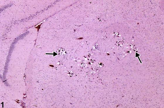
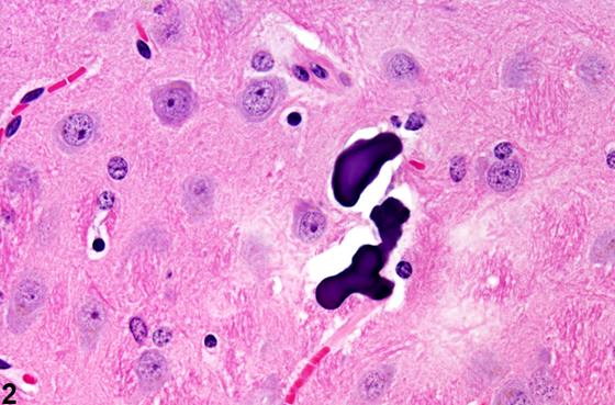
中枢神经系统(CNS)的矿化(mineralization)包括偶然发现的多灶性微小矿物质沉积,或坏死后发生的继发性营养不良性钙化(dystrophic calcification)。Figure 1、Figure 2 分别显示了大鼠和小鼠丘脑(thalamus)中偶然发现的多灶性微小矿物质沉积。丘脑内的分布通常为双侧性,但不一定对称。该病变在 B6C3F1 小鼠中比在大鼠中更常见。仔细观察时,矿物质颗粒常具有多层结构,在神经病理学上称为钙球(calcospherites)。它们似乎位于神经毡(neuropil)内,或常与毛细血管和小血管的内皮基底膜相关联。然而,即使血管受累且管腔狭窄,也罕见发现因其存在而导致丘脑微梗死的证据。该病变可能是钙代谢方面局灶性组织稳态异常的结果,在这些偶然性病例中,它不太可能代表继发于坏死的营养不良性矿化。近年来,老龄小鼠的丘脑病变已被认为与缺乏维生素 D 受体的小鼠相关。在所有 NTP 啮齿类研究组中均出现丘脑矿化(以钙球形式),证实了该现象的偶然性、特发性本质。当此类病变仅见于处理组,或在神经系统其他部位发现时,应考虑仔细评估是否存在营养不良性矿化、全身性高钙血症、维生素 D 过量或其他形式的钙稳态和代谢异常的证据。另一方面,继发于坏死的矿物质沉积归因于常见但复杂的营养不良性矿化过程。营养不良性矿化常表现为层状钙球,继发于多个神经部位的小簇状神经元坏死,如 Figure 3、Figure 4 所示。Figure 3 显示了小脑(cerebellum)中原颗粒细胞坏死部位矿化小体(箭头)的聚集。注意神经组织的丢失,表现为原内颗粒细胞占据的间隙,以及多个小的、深染的、圆形小体的存在。此类矿化灶的存在可能导致切片过程中组织破裂。Figure 3 所示的矿化灶不应与脑提取/修整过程中常从覆盖的颅骨碎片带入脑内的骨针人为假象或异位骨组织相混淆。Figure 4 中更高放大倍数显示的钙球呈圆形、常为同心圆、层状外观,是坏死脑组织矿化晚期的典型表现。钙球沉积于原小脑内颗粒细胞坏死区域。注意较大体部的同心层状结构(箭头),而较小体部中明显缺乏该结构。在这些病例中,以钙球形式沉积矿物质需要在坏死性损伤后数周才能形成。Figure 5 显示了毗邻梨状皮质(piriform cortex)的原坏死区域,伴有一个不常见的类骨质(osteoid)(箭头)和骨化(ossification)(箭头头)的反应性灶。一大片脑坏死区被几种类型矿化小体的进展所取代。右侧为一些较小的嗜碱性钙球,左侧为较大的坏死脑组织区域,已转化为粉红色透明物质构成的类骨质,以及更晚期的骨性化生(osseous metaplasia),其中致密嗜碱性矿物质和骨细胞明显可见。
诊断与分级建议(NTP 共识)
除非在对照组中缺失,丘脑矿化不解释为处理效应。如果与处理或剂量相关,则根据病变范围进行诊断和分级。营养不良性矿化应简单地诊断为矿化,无论残留病变谱(如钙球、类骨质或骨性化生)有何变化。此类变化应纳入描述中。诊断应包括具体部位,除非在整个脑区呈多灶性分布。严重程度分级基于病变范围。存在并发病变时,通常诊断最严重的病变。其他并发病变如有必要可根据严重程度单独诊断。
图谱
英文原文
展开 NTP 官方英文描述与建议
Comment:
Mineralization in the CNS comprises either incidental multifocal microscopic deposits of mineral or secondary dystrophic calcification occurring after necrosis. Figure 1 and Figure 2 show incidental multifocal microscopic deposits of mineral in the thalamus of rat and mouse, respectively. The thalamic distribution is usually bilateral but not necessarily symmetrical. It is a more common finding in B6C3F1 mice than in rats. The particles of mineral are often found, on close inspection, to have a multilamellar structure and are referred to neuropathologically as calcospherites. They appear to be located in the neuropil, or commonly associated with the intimal basement membrane of capillaries and small blood vessels. However, it is unusual to find any evidence of thalamic microinfarcts as a result of their presence, even when vessels are affected and the lumen is narrowed. The lesion is probably the result of focally abnormal tissue homeostasis with respect to calcium metabolism, and in these incidental cases, it is unlikely to represent dystrophic mineralization secondary to necrosis. More recently, thalamic lesions in aging mice have been associated with mice lacking vitamin D receptor. The occurrence of thalamic mineralization as calcospherites in all NTP rodent study groups confirms the incidental idiopathic nature of this phenomenon. When such lesions are seen in treated groups only, or when noted at other sites in the nervous system, careful evaluation for evidence of dystrophic mineralization, systemic hypercalcemia, vitamin D excess, or other forms of abnormal calcium homeostasis and metabolism should be considered. On the other hand, mineral deposits, secondary to necrosis, are attributed to the commonly observed but complex process of dystrophic mineralization. Dystrophic mineralization is often seen as lamellar calcospherites found secondary to small clustered regions of neuronal necrosis at various neural sites, such as depicted in Figure 3 and Figure 4 . Figure 3 shows the aggregation of mineralized bodies (arrow) in sites of former granule cell necrosis in the cerebellum. Note the loss of neural tissue represented by spaces formerly occupied by internal granule cells, and the presence of multiple small, dark, round bodies. The presence of such mineralized foci may lead to disruption of the tissue during microtomy. The mineralized foci shown in Figure 3 should not be confused with the artifactual presence of boney spicules often carried into brain from overlying skull fragments during brain extraction/trimming or with heterotopic bone tissue. The round, often concentric, lamellar appearance of calcospherites, shown at higher magnification in Figure 4 , is typical of late stages of mineralization of necrotic brain tissue. Calcospherites are deposited in a region of former cerebellar internal granule cell necrosis. Note the concentric laminated structures (arrow) of the larger bodies that are apparently absent in smaller ones. The deposition of mineral in the form of calcospherites in these cases requires several weeks to form after the necrotizing insult. Figure 5 shows a region of prior necrosis adjacent to piriform cortex with an uncommon reactive focus of osteoid (arrow) and ossification (arrowhead). A large zone of brain necrosis is replaced by a progression of mineralized bodies of several types. To the right are some smaller basophilic calcospherites, and to the left are larger zones of necrotic brain tissue transformed into both osteoid, composed of pink hyaline material, and a more advanced stage of osseous metaplasia, where dense basophilic mineral and osteocytes are apparent.
Recommendation:
Mineralization in the thalamus is not interpreted as a treatment effect unless it is absent in controls. If treatment or dose related, the lesion is diagnosed and graded based on the extent of the lesion. Dystrophic mineralization should be diagnosed simply as mineralization regardless of the variation in the spectrum of residual lesions such as calcospherites, or osteoid or osseous metaplasia. Such variations should be included in the narrative. The diagnosis should include the subsite unless the incidence is multifocal across brain regions. Severity grading is based on the extent of the lesion. In the presence of concurrent lesions, the most severe lesion is typically diagnosed. Other concurrent lesions may be diagnosed separately, if warranted by the severity.
来源与引用
原始条目:https://ntp.niehs.nih.gov/atlas/nnl/nervous-system/brain/Mineralization
图谱总目录:NTP Nonneoplastic Lesion Atlas。 内容属美国联邦政府作品(公有领域),可引用;引用时请标注来源。
建议引用格式:NTP Nonneoplastic Lesion Atlas. Mineralization (brain). LabSmart 中文版:
https://www.labsmart.cn/ntp/brain-mineralization.html
中文图谱总页(可展开对照全部 595 条):https://www.labsmart.cn/ntp-atlas.html#brain-mineralization