坏死Necrosis · 脑 · 神经系统 · NTP ntp-0384
诊断要点:在NTP研究中,多灶性梗死和软化被诊断为脑,多部位,坏死。如果坏死为局灶性(focal),诊断应包括亚部位。
病变描述
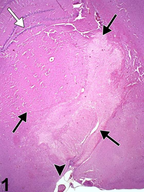
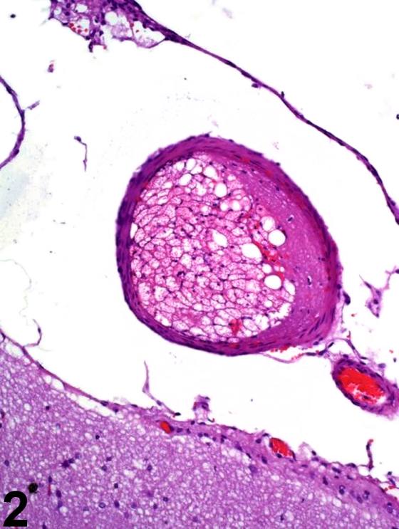
本节重点讨论由不同过程引起的脑坏死形态学及反应的性质。在NTP研究中,梗死被诊断为坏死,而“软化(malacia)”一词保留用于脑的大体病变。Figure 1显示了动脉栓子(箭头)所致急性血管闭塞的梗死效应。Figure 2中放大了纤维蛋白和脂肪栓子。Figure 1中白色箭头标识海马齿状回。注意提示受累区域早期坏死的苍白区(箭头),单侧累及大部分丘脑、下丘脑、大脑脚和视束。在缺血性脑病变中,神经元坏死的典型形态学标准在苏木精和伊红染色切片中可能需要超过24小时的存活时间才能显现。在Figure 1所示的时间点,病变边缘几乎没有,如果有的话,炎症细胞浸润的证据,表明梗死时间不足24小时。海马相对免受缺血影响。在大多数情况下,脑梗死的原因是偶然的,和/或归因于侵入性采样技术,如血液或骨髓采集,其中栓子进入静脉或动脉系统。Figure 3描绘了后丘(posterior colliculus)急性坏死的双侧对称性病变。还应注意外侧丘系核(nucleus of the lateral lemniscus)的坏死。边界清晰的苍白灶伴出血,双侧累及后丘核及外侧丘系核,类似于缺血性病变。与顶叶皮层一样,后丘负责听觉反射,具有高代谢率,特别易受干扰神经元能量代谢过程的影响。同样,Figure 4描绘了顶叶皮层1区(蓝色箭头)、丘脑(箭头)和压后皮层(白色箭头)的急性双侧坏死。脑区对毒性化合物效应的选择性易损性可能模仿缺血性坏死病变,应根据化合物暴露和临床病史与该类损伤相鉴别。神经病变的双侧对称性通常是毒性而非缺血性病因的有用指标。Figure 5描绘了脊髓背侧索中一种不寻常形式的区域性完全坏死(箭头)。这种类型的脊髓坏死被称为“脊髓出血(hematomyelia)”。它通常是邻近部位坏死或创伤性/肿瘤性脊髓压迫的结果,导致炎症核心的肿胀、出血和组织破坏在脊髓内向头侧和尾侧进展。Figure 6显示了伴有胶质增生和毛细血管增生的皮层梗死(箭头)。受累皮层通过缺血组织的轻微塌陷来识别,在病变的脑膜侧最为明显。即使在此低倍放大下,轻度皮层核固缩也显而易见。Figure 7显示了皮层梗死(箭头)的更晚期阶段,伴有胶质增生和巨噬细胞浸润。病变边界由胶质细胞反应界定,早期空洞形成正在整个区域进行。Figure 8描绘了已知持续时间的梗死形态。梗死72小时后,毛细血管核肥大(箭头)、早期巨噬细胞反应以及受累组织的一些碎裂显而易见。Figure 9显示了皮层梗死的较晚期阶段,此时组织丢失和皮层结构塌陷,在脑膜表面形成凹陷。这种皮层软膜下萎缩还伴有软脑膜间隙和空洞区域(箭头)的残留巨噬细胞。虽然在此苏木精和伊红切片中星形细胞肥大不明显,但胶质纤维酸性蛋白染色会显示与损伤持续时间相符的显著反应。Figure 10描绘了已知72小时持续时间的皮层梗死中的细胞反应(PB Little,个人观察)。注意毛细血管内皮的肥大和增生状态以及显著的巨噬细胞存在(箭头)。这些巨噬细胞已从驻留的小胶质细胞或血源性单核细胞分化而来,正在受累缺血区域吞噬脑脂质。它们被称为脂质吞噬细胞,或更常见地称为“格子细胞(gitter cells)”。它们还可通过365 nm波长下的自发荧光以及过碘酸雪夫染色或各种脂质染色(如Sudan III或油红O)的阳性染色进一步识别。Figure 11是严重嗅球坏死的例子,常被称为软化。该器官大部分实质丢失,伴有中央出血和坏死碎片(箭头)。软化是脑某一区域完全坏死的过程,描述大体可触及的大片受累脑坏死区域的质地。在NTP研究中,组织学上软化被诊断为坏死。显微镜下,病变中神经组织的所有成分均坏死。此处,过程已进展至坏死成分液化,仅剩无定形碎片和一些出血。最终,这样的区域注定成为充满脑脊液的囊肿。在晚期神经坏死区域,大部分可识别的组织丢失。重要的是将过程归因于坏死而非自溶,后者可能因缺乏存活神经组织而被提示。Figure 11所示邻近组织形态正常的存在有助于形成坏死的诊断。Figure 12勾勒了海马CA1区,其中存在缺血性坏死。微管相关蛋白免疫组化技术阴性勾勒出坏死区(箭头)。同样,如果研究中预期脑梗死,使用三苯基四氮唑有助于大体界定受累区域。此类特殊染色不用于常规一线评估,但了解它们在特殊情况下是有用的。
诊断与分级建议(NTP 共识)
在NTP研究中,多灶性梗死和软化被诊断为脑,多部位,坏死。如果坏死为局灶性(focal),诊断应包括亚部位。严重程度分级基于病变范围。当单个神经元受累时,诊断应为脑,神经元,坏死。坏死的相关特征应纳入叙述中。在晚期坏死病变中,还可见胶质增生和炎症、组织塌陷及囊肿形成时,通常诊断最严重的病变。其他并发病变,如果严重程度需要,可单独诊断。
图谱
英文原文
展开 NTP 官方英文描述与建议
Comment:
This section focuses on the morphology of brain necrosis caused by differing processes and the nature of the responses. In NTP studies, infarcts are diagnosed as necrosis, and the term “malacia” is reserved for gross lesions in the brain. Figure 1 identifies the infarctive effects of acute vascular occlusion by an arterial embolus (arrowhead). The fibrin and fat embolus is enlarged in Figure 2 . The hippocampal dentate gyrus is identified by a white arrow in Figure 1 . Note the pallor signifying early necrosis of the affected region (arrows) that involves much of the thalamus, hypothalamus, cerebral peduncle, and optic tract unilaterally. In ischemic brain lesions, the typical morphologic criteria of neuronal necrosis may take more than 24 hours of survival to manifest in hematoxylin and eosin sections. At the time point shown in Figure 1 , there is little, if any, evidence of inflammatory infiltrate at the margin of the lesion, indicating that the infarction is less than 24 hours old. The hippocampus is relatively spared from the ischemia. In most cases, the cause of brain infarction is incidental and/or attributable to invasive sampling techniques such as blood or marrow collection in which emboli gain access to the venous or arterial systems. Figure 3 depicts a bilaterally symmetrical lesion of acute necrosis of the posterior colliculus. Note also the necrosis of the nucleus of the lateral lemniscus. The well-defined foci of pallor, with hemorrhage, affecting the posterior collicular nucleus bilaterally, and the nucleus of the lateral lemniscus resemble those of ischemic lesions. Like the parietal cortex, the posterior colliculus, subserving auditory reflexes, has a high metabolic rate and is particularly vulnerable to processes that interfere with neuronal energy metabolism. Similarly, Figure 4 depicts acute bilateral necrosis in the parietal cortex area 1 (blue arrow), thalamus (arrowhead), and retrosplenial cortex (white arrow). Selective vulnerability of brain regions to the effects of toxic compounds may imitate the lesions of ischemic necrosis and should be differentiated from insults of that type based on compound exposure and clinical history. Bilateral symmetry of neural lesions is generally a useful indicator of toxic, rather than ischemic, etiology. Figure 5 depicts an unusual form of total regional necrosis (arrow) of the spinal cord in the dorsal spinal funiculi. This type of spinal cord necrosis is referred to as “hematomyelia.” It is usually the result of necrosis or traumatic/neoplastic spinal compression at an adjacent site with resulting propagation of an inflammatory core of swelling, hemorrhage, and tissue disruption that progresses anteriorly and posteriorly within the spinal cord. Figure 6 shows a cortical infarct with gliosis and capillary hyperplasia (arrow). The affected cortex is identified by minimal collapse of ischemic tissue, most noticeable at the meningeal extent of the lesion. Even at this low magnification, a mild degree of cortical nuclear pyknosis is apparent. In Figure 7 , a more advanced stage of cortical infarction (arrows) is shown, with gliosis and macrophage infiltration. The borders of the lesion are defined by the glial cell response, and early cavitation is proceeding throughout. Figure 8 depicts the morphology of an infarct of known duration. At 72 hours after infarction, the capillary nuclear hypertrophy (arrow), early macrophage response, and some fragmentation of the affected tissue are evident. Depicted in Figure 9 is a later stage of cortical infarction where there is loss of tissue and collapse of the cortical structure, creating a depression at the meningeal surface. This subpial atrophy of the cortex is also accompanied by residual macrophages in the leptomeningeal spaces and cavitated areas (arrows). While astrocytic hypertrophy is not apparent in this hematoxylin and eosin section, a glial fibrillary acidic protein stain would demonstrate a significant response coinciding with the duration of the insult. Figure 10 depicts the cellular responses in cortical infarction of known 72 hour duration (PB Little, personal observation). Note the hypertrophic and hyperplastic state of the capillary endothelium and the prominent macrophage presence (arrows). These macrophages have differentiated from resident microglial cells or blood-borne monocytic cells and are in the process of phagocytosis of brain lipid in the affected ischemic region. They are referred to as lipid phagocytes or, more commonly, as “gitter cells.” They can be further identified by their autofluorescence at 365 nm wavelength and by their positive staining with periodic acid Schiff or various lipid stains such as Sudan III or oil red O. Figure 11 is an example of severe olfactory bulb necrosis that has often been referred to as malacia. There is loss of much of the substance of the organ accompanied by central hemorrhage and necrotic debris (arrows). Malacia is a process of total necrosis of a region of brain and describes the gross palpable texture of the large affected necrotic region of brain. Histologically, in NTP studies, malacia is histologically diagnosed as necrosis. Microscopically, all elements of the neural tissue in a lesion are necrotic. Here, the process has advanced to liquefaction of necrotic elements, leaving little but amorphous debris and some hemorrhage. Ultimately, such an area is destined to become a cyst filled with cerebrospinal fluid. In regions of advanced neural necrosis, much of the recognizable tissue is lost. It is important to ascribe the process to necrosis rather than autolysis, which the lack of viable neural tissue may suggest. The presence of normal morphology of adjacent tissue as shown in Figure 11 is helpful in forming a diagnosis of necrosis. Figure 12 outlines the CA1 region in the hippocampus in which there is ischemic necrosis. Microtubule-associated protein immunohistochemical technique negatively outlines the necrotic zone (arrows). Similarly, if cerebral infarction is anticipated in a study, the use of triphenyltetrazolium is valuable to grossly define the region of involvement. Such special stains are not used in routine first-tier evaluation, but it is useful to be aware of them for special circumstances.
Recommendation:
In NTP studies, multifocal infarction and malacia are diagnosed as Brain, Multiple sites, Necrosis. If necrosis is focal, the diagnosis should include the subsite. Severity grading is based on the extent of the lesion. When individual neurons are affected, the diagnosis should be Brain, Neuron, Necrosis. Pertinent features of the necrosis should be included in the narrative. In advanced lesions of necrosis, where gliosis and inflammation, tissue collapse, and cyst formation are also seen, lesions with the most severity are typically diagnosed. Other concurrent lesions may be diagnosed separately, if warranted by the severity.
来源与引用
原始条目:https://ntp.niehs.nih.gov/atlas/nnl/nervous-system/brain/Necrosis
图谱总目录:NTP Nonneoplastic Lesion Atlas。 内容属美国联邦政府作品(公有领域),可引用;引用时请标注来源。
建议引用格式:NTP Nonneoplastic Lesion Atlas. Necrosis (brain). LabSmart 中文版:
https://www.labsmart.cn/ntp/brain-necrosis.html
中文图谱总页(可展开对照全部 595 条):https://www.labsmart.cn/ntp-atlas.html#brain-necrosis