小胶质细胞增生Microgliosis · 脑 · 神经系统 · NTP ntp-0385
诊断要点:当反应细胞以具有上述及图示典型小胶质细胞特征为主时,应使用“小胶质细胞增生(microgliosis)”一词。
病变描述
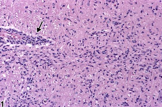
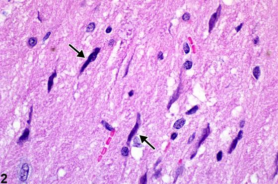
本节讨论脑内主要反应性细胞的细胞反应:小胶质细胞(microglia)、毛细血管内皮细胞(capillary endothelial cells)和星形胶质细胞(astrocytes)。小胶质细胞是早期反应细胞,在损伤部位及其周围12–24小时内即可显现。其向可识别巨噬细胞的演变在48小时内变得明显。作为巨噬细胞,其胞质增多,脂质降解产物可通过高碘酸-雪夫试剂(periodic acid Schiff reagent)及365 nm波长下的自发荧光检测到。此类吞噬细胞称为脂质吞噬细胞,或更常称为“泡沫细胞(gitter cells)”。Figure 1 显示炎症性单核细胞血管周围套(箭头)邻近的小胶质细胞增生。注意典型小胶质细胞众多细长、不规则的细胞核。该图像显示脑对损伤的反应,即通过激活驻留小胶质细胞实现。这些中胚层细胞在神经发育的早期胚胎阶段进入脑内,具有巨噬细胞能力。在非活跃状态下,它们分布于整个脑内,因其无特征的圆形细胞核及缺乏可见胞质而难以识别。当因脑损伤的存在而被激活时,它们变为可运动并迁移至损伤部位。在迁移过程中,细胞核变得细长且形状不规则,称为“杆状细胞(rod cells)”。通过这种细胞核形态,它们易于被检测到(如本图所示),分布于远离血管周围单核细胞浸润处。除驻留小胶质细胞外,血液和骨髓来源的单核细胞穿过内皮迁移至损伤部位,在那里它们也承担类似驻留小胶质细胞的吞噬功能。这解释了Figure 1(箭头)所示损伤脑组织邻近单核细胞一定程度血管周围套形成的原因。这些血源性单核细胞的外观和功能据知与驻留小胶质细胞无异。检测脑内这些细胞特别有用的免疫组化染色是Iba-1(离子化钙结合衔接分子1)抗体。Figure 2 显示小胶质细胞(杆状细胞)激活的细长不规则细胞核(箭头)。在高倍放大下显示,这些激活的小胶质细胞在趋化刺激下向神经损伤区域迁移。无法确定这些细胞是驻留小胶质细胞还是来源于经血流携带至神经组织的浸润性单核细胞。Figure 3 描绘了大脑皮质中小胶质细胞的Iba-1免疫组化检测。注意棕色染色的细胞及其突起广泛的放射状、横断面和纵断面(箭头)。Iba-1特异性表达于巨噬细胞和小胶质细胞,并在细胞激活过程中上调。Figure 4 显示既往损伤部位的反应性胶质增生和毛细血管增生(箭头)。毛细血管对邻近组织的损伤反应迅速,内皮细胞核肥大在24小时内即可明显。对病理学家而言,在低倍镜扫描脑组织时,这些毛细血管变化往往是某区域损伤的首个指征。本图中所见的反应性胶质增生很可能是浸润性血液单核细胞与驻留小胶质细胞的混合。尚无细胞分化为可识别的巨噬细胞。因此该病变可能不足48小时。使用层粘连蛋白(laminin)和血管性血友病因子(von Willebrand factor)进行免疫组化可增强对内皮反应的选择性研究。Figure 5 显示壳核(putamen)损伤的混合性胶质增生反应,由反应性星形胶质细胞、巨噬细胞、小胶质细胞和单核细胞组成。明显可见该区域有核细胞显著增多。在此放大倍数下,仅用苏木精-伊红(H&E)染色,无法识别构成该细胞群体的特定细胞类型。在许多研究中,仅需将反应识别为混合性胶质细胞型即可,其中包括星形胶质细胞、小胶质细胞和血源性单核细胞和/或已分化为吞噬细胞的巨噬细胞。在需要特异性识别浸润细胞类型的情况下,则需要特殊染色和免疫组化。正常星形胶质细胞,包括原浆型和纤维型,弥漫分布于神经系统中,其功能包括稳定结构、维持神经环境、神经元能量过程及血脑屏障。脑内正常星形胶质细胞群体的密度在软脑膜(pia mater)正下方的胶质界膜(glia limitans)以及血管周围增高。肥胖型星形胶质细胞增生(gemistocytic astrocytosis)是许多形式神经损伤进行性反应的组成部分,有助于发现细微脑损伤,并可大致判断现存病变的时间顺序。Figure 6 在高倍放大下显示H&E染色的愈合中神经损伤部位的反应性肥胖型星形胶质细胞增生。注意这些反应性星形胶质细胞典型的偏位核和显著的嗜酸性胞质(箭头),以及较少见的微小双核形式(箭头)。此外,正常的放射状突起变大且广泛。在此例中,其中一个反应性星形胶质细胞出现罕见的双核化形式,提示反应中不仅存在肥大,还存在增生。肥胖型星形胶质细胞的出现是判断脑病变时间的有用指标,因为在H&E切片上,损伤后12–14天之前其很少充分发育。使用针对胶质纤维酸性蛋白(glial fibrillary acidic protein,GFAP)的免疫组化技术——该蛋白构成星形胶质细胞中间丝——可检测反应的最早阶段。这大约在损伤后7天开始,使任何损伤部位更易识别,即使H&E切片上几乎看不到什么。因此,GFAP是细微脑损伤及相关星形胶质细胞反应非常有用、敏感的指标。虽然GFAP免疫反应性在神经病理学中广泛用于识别和定量星形胶质细胞,但其他一些细胞类型如室管膜细胞(ependymal cells)也可能表达低水平的该中间丝。
诊断与分级建议(NTP 共识)
当反应细胞以具有上述及图示典型小胶质细胞特征为主时,应使用“小胶质细胞增生(microgliosis)”一词。当反应细胞的性质不明显,且无识别细胞的特殊技术可用时,则适合使用术语胶质增生,NOS(未另行规定)。肥胖型星形胶质细胞增生存在时即作出诊断。在局灶性病变中,应注明神经解剖亚部位。若病变见于多个部位,应诊断为脑,小胶质细胞增生。在所有病例中,病变应进行分级。存在并发病变时,通常诊断严重程度最高的病变。其他并发病变若严重程度需要,可单独诊断。
图谱
英文原文
展开 NTP 官方英文描述与建议
Comment:
This section deals with the cellular responses of prominent reactive cells in the brain: microglia, capillary endothelial cells, and astrocytes. The microglia are early-responding cells and are apparent in and around an injured site within 12–24 hours. Their evolution to recognizable macrophages is apparent within 48 hours. As macrophages, they have increased cytoplasm, and products of lipid degradation can be detected with periodic acid Schiff reagent and by autofluorescence at 365 nm wavelength. Such phagocytic cells are referred to as lipid phagocytes or, more commonly, “gitter cells.” Figure 1 depicts microgliosis adjacent to an inflammatory mononuclear perivascular cuff (arrow). Note the many elongated, irregular nuclei of typical microglial cells. This image depicts the response of brain to injury by the activation of resident microglial cells. These mesodermal cells enter the brain at an early embryological stage of neural development and have macrophage capability. In the inactive state, they are distributed throughout the brain and are difficult to identify because of their nondiscriminate round nuclear character and lack of visible cytoplasm. When they are activated by the presence of brain injury, they become motile and migrate to the site of injury. In the process of migration, nuclei become elongated and irregular in shape and are referred to as “rod cells.” By this nuclear morphology, they are easily detected (as shown in this image), distributed distant from the perivascular monocytic infiltrate. As well as resident microglia, mononuclear cells of blood and bone marrow origin cross the endothelium and migrate to the injured site, where they also take up phagocytic functions like those of resident microglia. This accounts for the presence of some degree of perivascular cuffing by monocytes adjacent to injured brain tissue, as shown in Figure 1 (arrow). The appearance and function of these blood borne monocytes are not known to be different from those of resident microglia. A particularly useful immunohistochemical stain to detect these cells in brain is the Iba-1 (ionized calcium binding adaptor molecule 1) antibody. Figure 2 shows activated elongated irregular nuclei (arrows) of microglial cells (rod cells). Shown at high magnification, these activated microglial cells are migrating under chemotactic stimuli to a region of neural injury. It is not possible to say whether these cells are resident microglia or those derived from infiltrating monocytic cells carried to the nervous tissue by the blood stream. Depicted in Figure 3 is the Iba-1 immunohistochemical detection of microglia in cerebral cortex. Note the brown-staining cells and extensive radiating, cross, and longitudinal sections of their processes (arrows). Iba-1 is specifically expressed in macrophages and microglia and is upregulated during cellular activation. Figure 4 shows reactive gliosis and capillary hyperplasia (arrow) in a site of former injury. Capillaries are quickly responsive to injury of adjacent tissue, and endothelial nuclear hypertrophy is evident within 24 hours. To the pathologist these capillary changes are often, upon low-magnification scanning of brain tissue, the first indication of damage in a region. The reactive gliosis seen in this image is likely a mixture of infiltrating blood monocytes and resident microglial cells. None have yet differentiated to recognizable macrophages. The lesion is therefore probably less than 48 hours old. Selective study of endothelial responses may be enhanced by the use of immunohistochemistry using laminin and von Willebrand factor. Figure 5 displays a mixed gliotic response to injury of the putamen, consisting of reactive astrocytes, macrophages, microglia, and mononuclear cells. It is apparent that there is a significant increase in nucleated cells in the region. At this magnification, and with hematoxylin and eosin (H&E) staining alone, it is not possible to identify the specific cell types that make up the population. In many studies, it is only relevant to identify the response as being of mixed glial type, which would include astrocytes, microglial cells, and monocytic cells of blood origin and/or macrophages if they have already differentiated to phagocytic cells. In those cases where specific identification of the types of infiltrating cells is necessary, special stains and immunohistochemistry would be required. Normal astrocytes, protoplasmic and fibrillary, are diffusely distributed in the nervous system, where they serve, among other functions, to stabilize structure and maintain the neural milieu, neuronal energy processes, and the blood brain barrier. The density of the normal astrocyte population in the brain is increased in the glia limitans just beneath the pia mater, and also around blood vessels.Gemistocytic astrocytosis is an integral part of the progressive response to many forms of neural injury and is helpful in uncovering subtle brain injury, as well as giving an approximation of the chronology of the lesions present. Figure 6 shows at high magnification reactive gemistocytic astrocytosis in a site of healing neural injury stained with H&E. Note the typical eccentric nucleus and prominent eosinophilic cytoplasm of these reactive astrocytes (arrowheads) and the less frequent micro-binucleate form (arrow). In addition, the normal radiating processes become enlarged and extensive. In this example, there is a rare form of binucleation of one of the reactive astrocytes, signaling the presence not only of hypertrophy but also of hyperplasia in the response. The appearance of gemistocytic astrocytes is a useful indicator of the age of a brain lesion since it is seldom well developed on H&E sections until 12–14 days after injury. The use of the immunohistochemical technique for glial fibrillary acidic protein (GFAP), which constitutes the astrocytic intermediate filaments, will detect the earliest stages of the response. This commences at approximately 7 days postinjury and makes any site of injury more recognizable even if there is little to see with H&E sections. In this way, GFAP is a very useful, sensitive indicator of subtle brain injury and the associated astrocytic responses. While GFAP immunoreactivity is used extensively in neuropathology to identify and quantify astrocytes, some other cell types such as ependymal cells may also express a low level of this intermediate filament.
Recommendation:
The term “microgliosis” should be used when the preponderance of reactive cells have the typical microglial features mentioned and illustrated above. When the nature of the reactive cells is not clearly apparent, and special techniques to identify the cells are not available, then it is appropriate to use the term Gliosis, NOS (not otherwise specified). Gemistocytic astrocytosis is diagnosed, when present. In focal lesions, the neuroanatomic subsite should be noted. If the lesion is noted in multiple sites, it should be diagnosed as Brain, Microgliosis. In all cases, the lesion should be graded. In the presence of concurrent lesions, lesions with the most severity are typically diagnosed. Other concurrent lesions may be diagnosed separately, if warranted by the severity.
来源与引用
原始条目:https://ntp.niehs.nih.gov/atlas/nnl/nervous-system/brain/Microgliosis
图谱总目录:NTP Nonneoplastic Lesion Atlas。 内容属美国联邦政府作品(公有领域),可引用;引用时请标注来源。
建议引用格式:NTP Nonneoplastic Lesion Atlas. Microgliosis (brain). LabSmart 中文版:
https://www.labsmart.cn/ntp/brain-microgliosis.html
中文图谱总页(可展开对照全部 595 条):https://www.labsmart.cn/ntp-atlas.html#brain-microgliosis