胆固醇裂隙Cholesterol Clefts · 脑 · 神经系统 · NTP ntp-0392
诊断要点:胆固醇裂隙存在时,在NTP研究中必须作出诊断并记录亚部位。存在并发病变时,通常诊断严重程度最高的病变。
病变描述
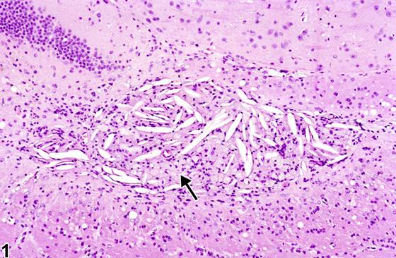
Figure 1(箭头)展示了一个成熟的退行性病灶,由胶质增生(gliosis)、巨噬细胞和结晶状胆固醇裂隙(cholesterol clefts)组成。这一小的消退性退行性病变毗邻海马齿状回。最显著的特征是曾由胆固醇结晶占据的空的长形针状空隙,通常称为胆固醇裂隙。溶剂组织处理已去除胆固醇。与胆固醇裂隙相关的是许多混合性胶质细胞和巨噬细胞。此类过程通常由脑坏死伴髓鞘蛋白脂质复合物释放所致,或源于慢性炎症过程中对外渗血液或降解细胞的吞噬作用。溶解细胞膜产生的降解脂质导致游离胆固醇和三酰甘油及其他化合物在细胞内蓄积。当巨噬细胞负载这些后述化合物且通常防止过量游离胆固醇蓄积的细胞安全机制失效时,即发生巨噬细胞凋亡。巨噬细胞释放游离胆固醇,后者在邻近组织中沉淀为结晶。这些胆固醇结晶在组织中存留很长时间,常为终生。脑组织中胆固醇结晶的显著外观通常应被识别为对既存脑坏死、慢性炎症或出血的反应的组成部分。
诊断与分级建议(NTP 共识)
胆固醇裂隙存在时,在NTP研究中必须作出诊断并记录亚部位。存在并发病变时,通常诊断严重程度最高的病变。其他并发病变若严重程度允许,可单独诊断。
图谱
英文原文
展开 NTP 官方英文描述与建议
Comment:
A mature degenerate focus is represented in Figure 1 (arrow) consisting of gliosis, macrophages, and crystalline-shaped cholesterol clefts. This small resolving degenerative lesion is adjacent to the dentate gyrus of the hippocampus. The most striking feature is the empty elongated spicular spaces formerly occupied by cholesterol crystals, often referred to as cholesterol clefts. Tissue processing with solvent has removed the cholesterol. Associated with cholesterol clefts are many mixed glial cells and macrophages. This type of process is usually the result of brain necrosis with liberation of myelin proteolipid complexes or from phagocytosis of extravasated blood or degraded cells in chronic inflammatory processes. The degraded lipid from lysed cell membranes results in intracellular accumulation of free cholesterol and triacylglycerol, along with other compounds. Apoptosis of macrophages results when they are loaded with these latter compounds and the usual cellular safety mechanisms preventing excess free cholesterol accumulation fail. The macrophages release free cholesterol, which precipitate as crystals in the adjacent tissue. These cholesterol crystals remain in the tissue for prolonged, often lifelong, periods. The striking appearance of cholesterol crystals in brain tissue should be recognized usually as a component of the response to pre-existing brain necrosis, chronic inflammation, or hemorrhage.
Recommendation:
Cholesterol clefts, when present, must be diagnosed in NTP studies and the subsite recorded. In the presence of concurrent lesions, lesions with the most severity are typically diagnosed. Other concurrent lesions may be diagnosed separately, if warranted by the severity.
来源与引用
原始条目:https://ntp.niehs.nih.gov/atlas/nnl/nervous-system/brain/CholesterolClefts
图谱总目录:NTP Nonneoplastic Lesion Atlas。 内容属美国联邦政府作品(公有领域),可引用;引用时请标注来源。
建议引用格式:NTP Nonneoplastic Lesion Atlas. Cholesterol Clefts (brain). LabSmart 中文版:
https://www.labsmart.cn/ntp/brain-cholesterol-clefts.html
中文图谱总页(可展开对照全部 595 条):https://www.labsmart.cn/ntp-atlas.html#brain-cholesterol-clefts