表皮样囊肿Epidermoid Cyst · 脑 · 神经系统 · NTP ntp-0396
诊断要点:尽管表皮样囊肿通常被认为是背景病变,但在NTP研究中应连同亚部位一起诊断。表皮样囊肿无需分级。
病变描述
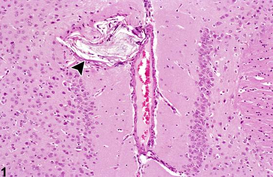
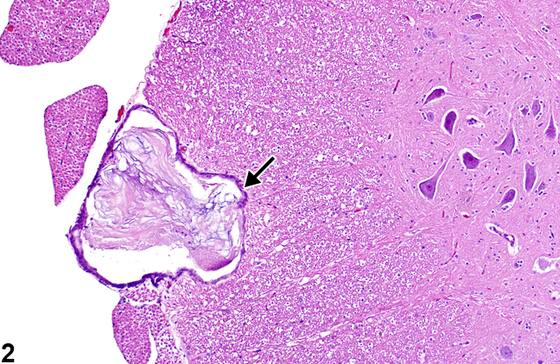
表皮样囊肿(epidermoid cyst)是良性错构瘤性结构,从其位置和大小来看,可能干扰正常神经功能,但通常临床上无症状,因为其体积缓慢增大,使神经系统能够对占位性病变的存在进行适应和代偿。在图1中,发育不良的表皮样囊肿位于背侧额顶中线位置的扣带皮质回。此类表皮样囊肿被认为起源于表皮组织的胚胎残余,当神经管与上覆神经板分离时,这些残余与内陷的神经管保持密切关联。囊肿形成过程中压迫皮质,具有薄层表皮衬里,通常为复层鳞状上皮,核心处有少量角蛋白碎屑。注意双侧胼胝体上回(induseum griseum),这是一个紧凑的正常神经元细胞区域,属于与海马中继相关的边缘系统的一部分。图2描绘了一个位于腰段腹侧脊髓索的蛛网膜下表皮样囊肿的更晚期发展。其外侧毗邻脊神经根,内侧毗邻腹侧脊髓灰质柱。索组织受到广泛压迫显而易见,但未在该动物中检测到临床症状。图3显示了位于软脑膜下方的偶发性皮质蛛网膜下表皮样囊肿的位置。存在对下方皮质的压迫以及邻近核团神经元的丢失。角蛋白核心中央部分存在营养不良性矿化。在图4中,这个大型偶发性表皮样囊肿位于间脑上方的中线位置,压迫邻近的海马齿状回和缰核结构。注意该囊肿细胞丰富且角化程度相对较差。在图5中,表皮样囊肿局限于脊髓神经根(radicle),其进行性增大已完全破坏神经纤维,仅留下神经外膜和神经束膜完整。
诊断与分级建议(NTP 共识)
尽管表皮样囊肿通常被认为是背景病变,但在NTP研究中应连同亚部位一起诊断。表皮样囊肿无需分级。
图谱
英文原文
展开 NTP 官方英文描述与建议
Comment:
Epidermoid cysts are benign hamartomatous structures, which, from their location and size, may interfere with normal neural functions but are usually clinically silent since the slow increase in size allows for adaptation and compensation by the nervous system to the presence of the space-occupying mass. In Figure 1 , the poorly developed epidermoid cyst is located in the cingulate cortical gyrus at the dorsal frontoparietal midline location. Such epidermoid cysts are considered to arise from embryonic rests of epidermal tissue that remain closely associated with the infolding neural tube when it separates from the overlying neural plate. The cyst, as it forms, compresses the cortex and has a thin epidermal, often stratified squamous epithelial lining and modest amount of keratin debris at the core. Note the bilateral induseum griseum, a compact normal region of neuronal cells, which is part of the limbic system associated with hippocampal relays. Figure 2 depicts a more advanced development of a sub-leptomeningeal epidermoid cyst located in the lumbar ventral spinal funiculus. Adjacent laterally are spinal nerve roots, and adjacent internally is the ventral spinal gray matter column. The extensive compression of the funicular tissue is apparent, but no clinical signs were detected in the animal. Figure 3 shows the location of an incidental cortical sub-leptomeningeal epidermoid cyst underlaid by the pia mater. There is compression of the underlying cortex and loss of neurons in the adjacent nucleus. Dystrophic mineralization of the central portion of the keratin core is present. In Figure 4 , this large incidental epidermoid cyst has a midline location above the diencephalon and compresses the adjacent hippocampal dentate gyrus and habenular nuclear structures. Note that the cyst is cellular and relatively poorly keratinized. In Figure 5 , the epidermoid cyst confines itself to a spinal radicle (nerve root), where its progressive enlargement has totally effaced the nerve fibers, leaving only the epineurium and perineurium intact.
Recommendation:
Although epidermoid cysts are generally considered background lesions, they should be diagnosed in NTP studies along with the subsite. Epidermoid cysts do not need to be graded.
来源与引用
原始条目:https://ntp.niehs.nih.gov/atlas/nnl/nervous-system/brain/EpidermoidCyst
图谱总目录:NTP Nonneoplastic Lesion Atlas。 内容属美国联邦政府作品(公有领域),可引用;引用时请标注来源。
建议引用格式:NTP Nonneoplastic Lesion Atlas. Epidermoid Cyst (brain). LabSmart 中文版:
https://www.labsmart.cn/ntp/brain-epidermoid-cyst.html
中文图谱总页(可展开对照全部 595 条):https://www.labsmart.cn/ntp-atlas.html#brain-epidermoid-cyst